By William Soriano, DC | Upper Cervical Chiropractor | Apex Chiropractic
The craniocervical junction, or CCJ, is the highly specialized region where the skull meets the upper cervical spine. It includes the occiput at the base of the skull, the atlas (C1), the axis (C2), their joints and ligaments, surrounding muscles, the upper spinal cord and lower brainstem, cerebrospinal fluid spaces, and important vascular structures. Few regions of the human body face such conflicting mechanical demands. The craniocervical junction must simultaneously:
- support the weight of the head
- permit a remarkably large range of movement
- protect the brainstem and upper spinal cord
- maintain stability around the dens
- accommodate the vertebral arteries
- provide continuous sensory information about head and neck position
- maintain an open passage between the cranial and spinal compartments
For this reason, the CCJ should not be thought of simply as “the top two bones in the neck.” It is an integrated biomechanical and neurological system. Modern radiological reviews similarly describe the craniocervical junction as an anatomically distinct osteoligamentous region with demands for both substantial mobility and protection of critical neural and vascular structures. 1
What Structures Make Up the Craniocervical Junction?
The principal bony structures are:
Occiput (C0) — specifically the portion of the skull surrounding the foramen magnum and the paired occipital condyles.
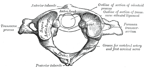
Atlas (C1) — the ring-shaped first cervical vertebra. Unlike the typical cervical vertebra, C1 has no conventional vertebral body or spinous process. Its lateral masses act as a cradle for the occipital condyles to support the skull.
Axis (C2) — distinguished by the dens, or odontoid process, which projects superiorly through the ring of C1 and acts as an important pivot for the atlas to rotate around.
Together they form two major joint complexes:
C0-C1: the atlanto-occipital joints
and
C1-C2: the atlantoaxial joints, including a medial joint around the dens and paired lateral joints.
These two motion segments are mechanically very different from the subaxial cervical spine below them.


Why the Upper Cervical Spine Is Different
From C2-C3 downward, cervical vertebrae are separated by intervertebral discs. There is no intervertebral disc between the occiput and C1 or between C1 and C2. Instead, movement and stability at the CCJ depend heavily on specialized joint geometry and an intricate ligamentous system. At C0-C1, the convex occipital condyles articulate with the concave superior articular surfaces of the atlas. Their geometry favors flexion and extension while relatively limiting axial rotation.
At C1-C2, the mechanical situation changes. The atlas rotates around the dens, while the lateral atlantoaxial articulations permit substantial three-dimensional movement.
Due to this, the atlantoaxial segment makes up the majority of cervical rotation. C1-C2 is a major contributor to cervical axial rotation. 2 That enormous mobility explains why C1-C2 depends so heavily on its ligamentous restraints.
The Ligamentous Stability System
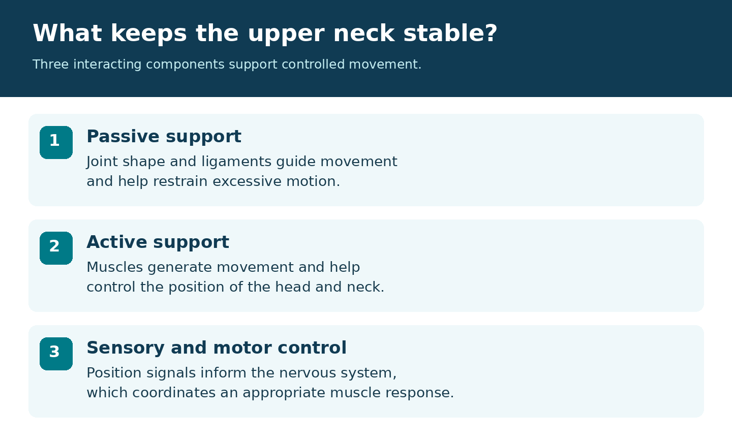
The craniocervical ligaments do not work independently. 3 Stability is produced by the interaction between:
- the transverse ligament of the atlas
- alar ligaments
- cruciform ligament complex
- tectorial membrane
- joint capsules
- atlanto-occipital and atlantoaxial membranes
- apical ligament
- several smaller accessory ligaments
- bony anatomy
- surrounding musculature
- neuromuscular control
The Transverse Ligament of the Atlas
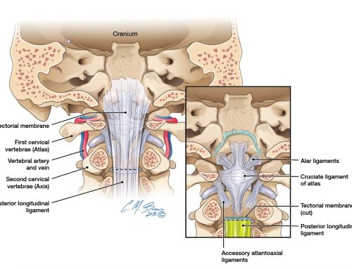
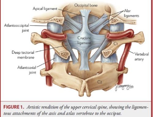
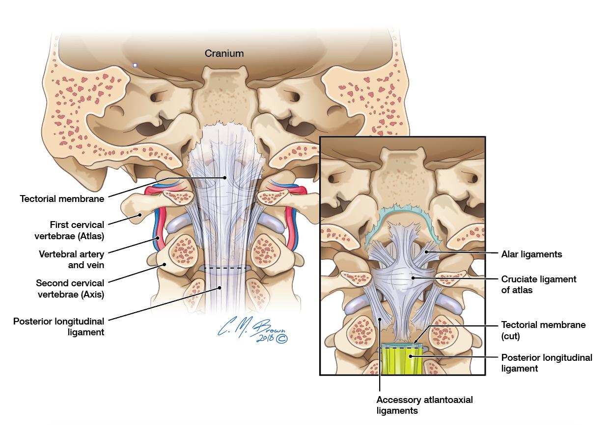
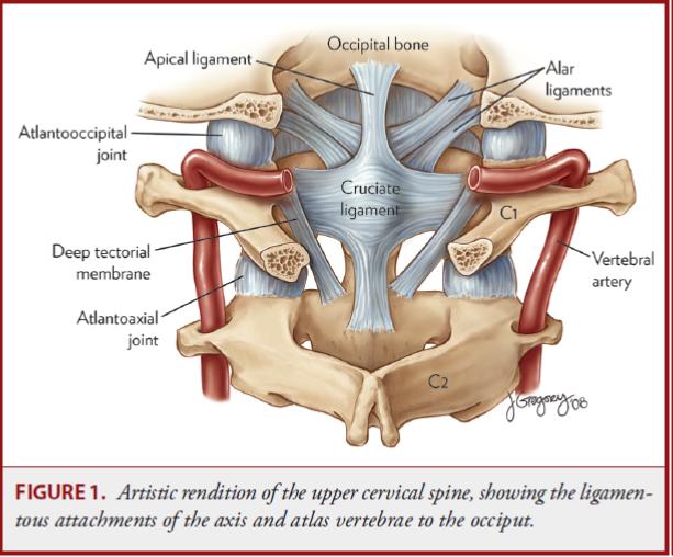
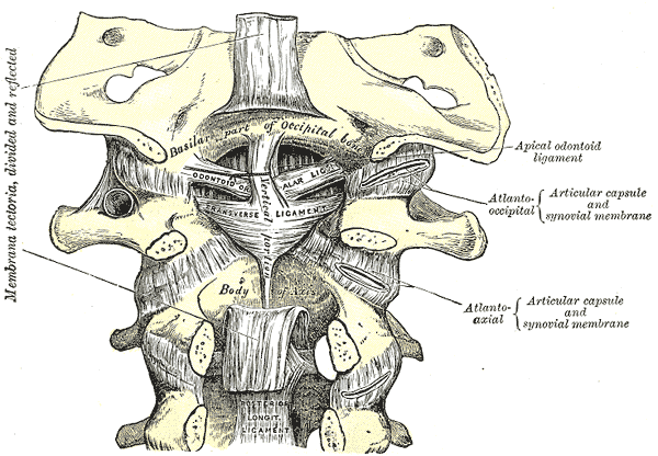
The transverse atlantal ligament passes behind the dens between the medial surfaces of the lateral masses of C1. Its primary mechanical function is to keep the dens closely approximated to the anterior arch of the atlas while still permitting C1 to rotate around C2. It is generally considered one of the most important stabilizing structures of the atlantoaxial articulation.
Disruption can permit abnormal anterior translation of C1 relative to C2 and significantly alter the relationship between the dens and the spinal canal. 1 The transverse ligament also forms the horizontal component of the cruciform ligament.
The Alar Ligaments
The paired alar ligaments extend primarily from the upper lateral dens toward the occipital region near the medial occipital condyles. Their orientation varies between individuals. Functionally, they are particularly important restraints to excessive axial rotation and lateral bending while also contributing to multiplanar stability.
Experimental studies demonstrate increased CCJ motion after alar-ligament sectioning, but the alar ligaments should not be treated as simple mechanical “stop cords.” Their tension changes during complex coupled movements, and they operate alongside the transverse ligament, capsules, tectorial membrane, bone, and musculature. 1 We discuss the structure in much greater detail in our comprehensive guide to the alar ligaments.
The Cruciform Ligament
The cruciform, or cruciate, ligament consists of the transverse ligament together with superior and inferior longitudinal bands. The longitudinal bands extend superiorly toward the occipital region and inferiorly toward C2. They contribute less mechanical restraint than the transverse component itself, which is why references to the “cruciform ligament” and “transverse ligament” should not be treated as mechanically equivalent. 1
The Tectorial Membrane
The tectorial membrane lies posterior to the dens and cruciform ligament and represents the cranial continuation of the posterior longitudinal ligament. Superiorly, it attaches to the clivus and blends intimately with the cranial dura. The tectorial membrane contributes to restraint of excessive motion alongside the other stabilizers. Its mechanical contribution depends on the position and loading of the craniocervical junction. 1
Its close relationship with the dura also makes it anatomically interesting beyond its purely ligamentous function.
The Joint Capsules and Smaller Ligaments
The atlanto-occipital and atlantoaxial joints are surrounded by capsular ligaments. Additional structures—including the apical ligament, accessory atlantoaxial ligaments, transverse occipital ligament, Barkow ligament and others—have been described anatomically. Some demonstrate considerable anatomical variability, and their individual contribution to stability is much smaller than that of the major ligamentous restraints.
For this reason, identifying an abnormality involving a minor ligament does not automatically establish clinically significant instability. 1
Craniocervical Biomechanics
The CCJ permits movement in all three anatomical planes:
Flexion and extension
Axial rotation
Lateral bending
These movements are not completely independent. Normal cervical motion involves coupled movement, meaning that rotation in one plane is frequently accompanied by smaller movements or translations in other planes.
C0-C1: Primarily Flexion and Extension
The geometry of the atlanto-occipital articulations favors flexion and extension. Rotation is comparatively limited. This is the familiar “yes” motion traditionally associated with C0-C1, although that description is an oversimplification because movement is three-dimensional rather than purely sagittal.
C1-C2: Primarily Axial Rotation
C1-C2 is the major rotational segment of the cervical spine. Measured upper-cervical rotation varies with joint position, age and testing method. Laboratory measurements of isolated structures should not be interpreted as a normal target for every patient. 4 This remarkable rotational capacity exists because of the shape of the atlantoaxial joints and the relationship between C1 and the dens. The cost of that mobility is greater dependence on ligamentous and muscular control.
The Muscular System of the CCJ
Deep beneath the larger posterior cervical musculature lies a specialized group known as the suboccipital muscles:
- rectus capitis posterior major
- rectus capitis posterior minor
- obliquus capitis superior
- obliquus capitis inferior
These small muscles contribute to extension, rotation, lateral movement, and fine control of head position. But viewing them merely as motors probably understates their importance.
The Upper Cervical Spine as a Sensory System
The cervical spine contains a sophisticated proprioceptive system. Proprioception is the nervous system’s ability to determine where a body part is located and how it is moving without having to see it. Research has demonstrated particularly high muscle-spindle content within the small deep cervical and suboccipital muscles. Muscle spindles appear to represent a major source of cervical proprioceptive information. 5
That information interacts centrally with visual and vestibular information involved in:
- head orientation
- postural control
- balance
- eye-head coordination
- spatial orientation
Cervical sensory information is integrated with visual and vestibular input. A disturbance in one source can affect the coordination of head movement and balance. 5 This helps explain why cervical sensorimotor dysfunction is increasingly investigated in patients experiencing neck pain, disequilibrium, and some forms of dizziness.
It does not, however, mean that dizziness automatically originates in the neck. Vestibular, neurological, cardiovascular, medication-related, metabolic, visual, and other causes must also be considered.
The Trigeminocervical Complex and Headache
Another important neurological relationship exists between the upper cervical spine and the trigeminal sensory system. Nociceptive afferents from structures innervated primarily by the upper cervical nerves converge with trigeminal nociceptive pathways in what is commonly called the trigeminocervical complex. This convergence provides a well-established neuroanatomical mechanism by which pain originating from cervical structures can be perceived in regions of the head. 6
Potential cervical pain generators include upper cervical joints, muscles, ligaments and other structures supplied by C1-C3. This is an important distinction:
The cervical spine can contribute to some headaches, but the presence of headache does not prove that the craniocervical junction is the cause.
Migraine, tension-type headache, vascular disorders, intracranial pathology and numerous other conditions can produce overlapping symptoms.
The Myodural Bridge Complex
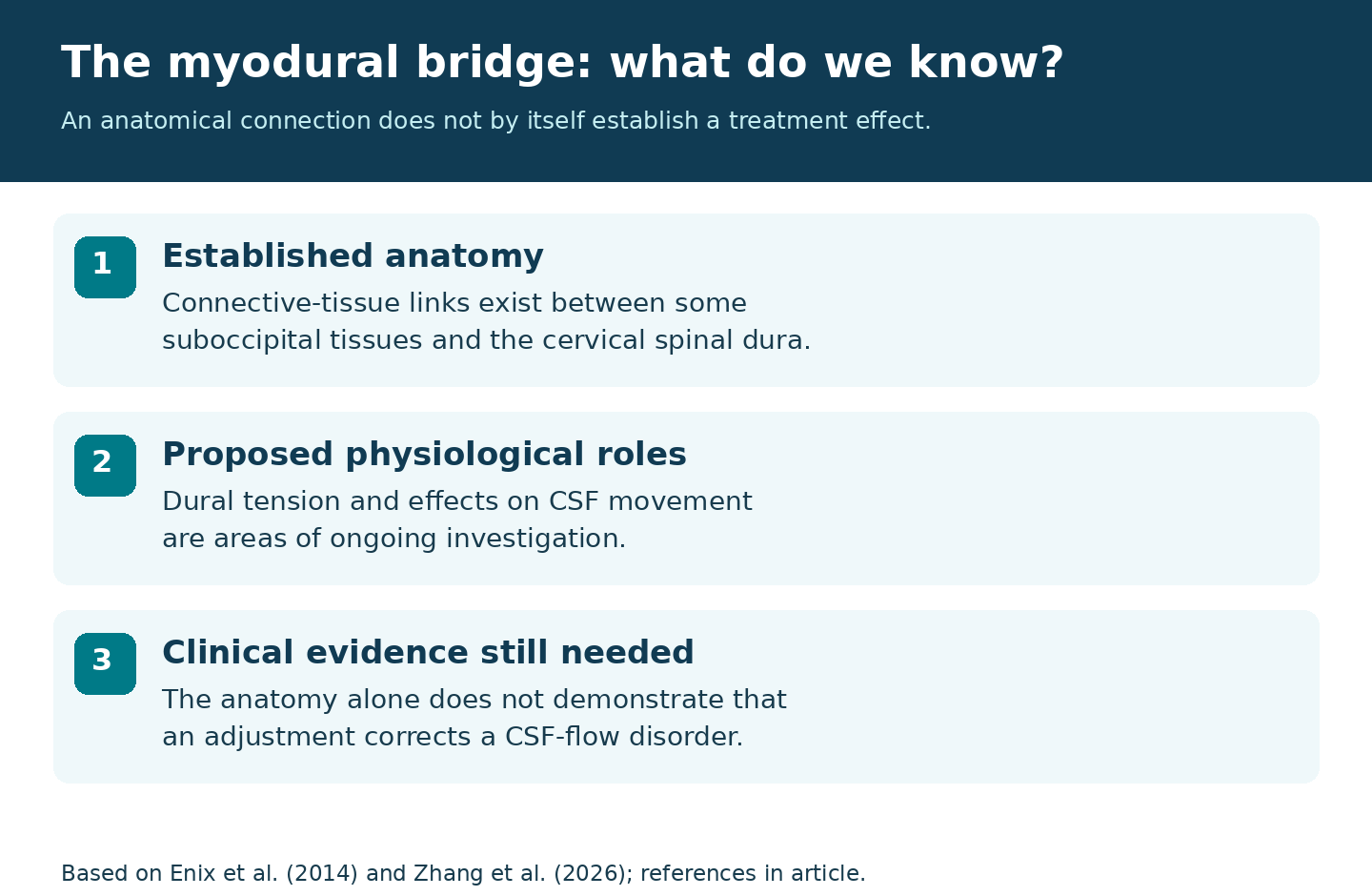
One of the more fascinating anatomical relationships at the CCJ is the myodural bridge complex. Fibrous connective-tissue structures link portions of the suboccipital musculature and related tissues to the cervical spinal dura, particularly through the posterior atlanto-occipital and atlantoaxial regions. Connections have been described involving:
- rectus capitis posterior minor
- rectus capitis posterior major
- obliquus capitis inferior
- the nuchal ligament
The anatomical existence of these connections is well established. 8 Proposed functions include maintaining dural tension during head movement, preventing dural infolding, contributing to proprioceptive signaling, and influencing cerebrospinal-fluid dynamics.
A comprehensive 2026 review concludes that mechanical force transmission through the myodural bridge is supported anatomically, while several broader physiological and clinical roles remain active areas of investigation. 7
The Brainstem and Upper Spinal Cord
The CCJ surrounds the transition between the medulla oblongata and cervical spinal cord. This region contains or lies near pathways responsible for an enormous range of functions, including:
- motor control
- somatosensory transmission
- balance and coordination
- autonomic regulation
- respiratory regulation
- lower cranial-nerve function
This does not mean that ordinary mechanical dysfunction of the upper cervical joints routinely compresses the brainstem. Actual brainstem or spinal-cord compression is a potentially serious neurological finding requiring appropriate medical evaluation and imaging. Conditions capable of compromising this region include severe trauma, congenital abnormalities, inflammatory disease, mass lesions, advanced instability, significant basilar invagination and some craniocervical malformations.
The Vertebral Arteries and the CCJ
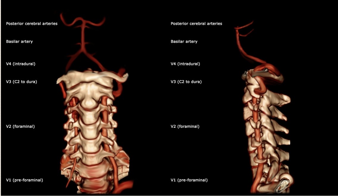
The vertebral arteries have an unusually complex course through the upper cervical region. In the upper cervical region, each vertebral artery ascends toward C1 and curves over the posterior arch of the atlas before piercing the dura. This mobile extracranial portion is commonly described as V3; the intradural segment is V4. Anatomical variants are important when interpreting this course.
This tortuous course provides mobility but also creates substantial anatomical variation. 9 The relationship is clinically important in:
- trauma
- congenital vascular variants
- surgery
- instrumentation
- vertebral-artery dissection or other vascular pathology
Normal neck movement should not be described as routinely cutting off vertebral blood flow. However, pathological anatomy, severe displacement, vascular abnormalities or traumatic injury can produce clinically important vascular consequences.
Cerebrospinal Fluid and the Foramen Magnum
The CCJ is also the gateway between the cranial and spinal cerebrospinal-fluid compartments. CSF circulates within the ventricular system and the subarachnoid spaces surrounding the brain and spinal cord. At the foramen magnum, CSF moves between the posterior fossa and cervical spinal subarachnoid space in a pulsatile manner influenced substantially by the cardiac cycle and respiration. Modern phase-contrast MRI can visualize and quantify aspects of this flow. 10
Normal flow is complex rather than a simple continuous downward stream. Research using phase-contrast MRI and computational fluid dynamics has demonstrated changing direction and velocity during the cardiac cycle, particularly around the foramen magnum and upper cervical canal. 11
There is experimental interest in whether suboccipital muscle contraction and myodural-bridge tension contribute additional mechanical energy to CSF movement. That hypothesis is intriguing but should remain distinguished from the much better-established cardiovascular and respiratory influences on CSF pulsatility. 7
Cerebellar Tonsils, Chiari, and the CCJ
The cerebellar tonsils lie near the foramen magnum, and their position varies between individuals. When the tonsils extend inferiorly beyond expected limits, they may be described as cerebellar tonsillar ectopia. A Chiari I malformation involves more than simply assigning significance to a single millimeter measurement; posterior-fossa anatomy, symptoms, neural structures, syringomyelia and CSF dynamics can all matter.
Phase-contrast MRI may demonstrate altered CSF flow in patients with Chiari I, particularly where crowding at the foramen magnum produces abnormal flow patterns. 10 Tonsillar position alone does not determine clinical significance.
Imaging the Craniocervical Junction
No single imaging modality answers every CCJ question.
Plain Radiography
Radiographs can demonstrate:
- gross alignment
- fractures in selected circumstances
- degenerative changes
- congenital anomalies
- certain relationships between C0, C1 and C2
- dynamic motion when appropriately indicated and performed
They provide limited direct evaluation of neural and ligamentous tissues.
Computed Tomography
CT provides excellent assessment of bone.
It is particularly useful for:
- fracture
- complex osseous anatomy
- occipital condyles
- C1 and C2
- congenital variants
- traumatic alignment
- preoperative assessment
In adults with acute cervical trauma who meet criteria for imaging, CT is commonly used to evaluate fractures and alignment. Imaging decisions in children require age-specific assessment. 12
Cone-Beam CT
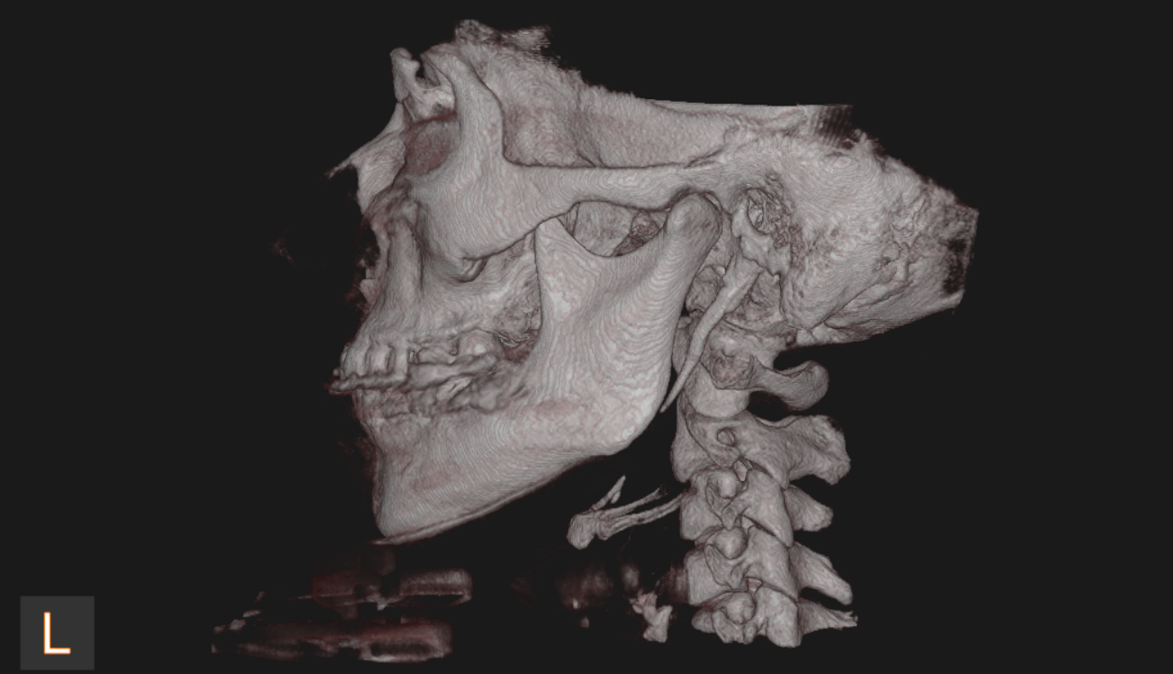
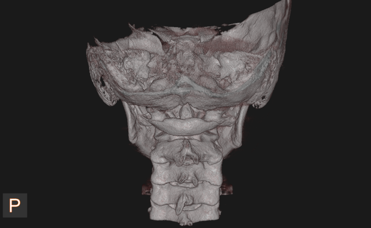
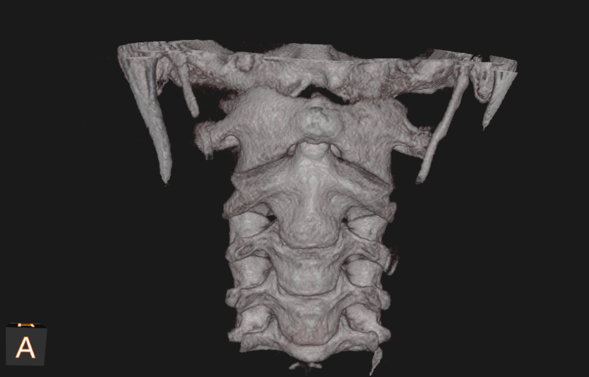
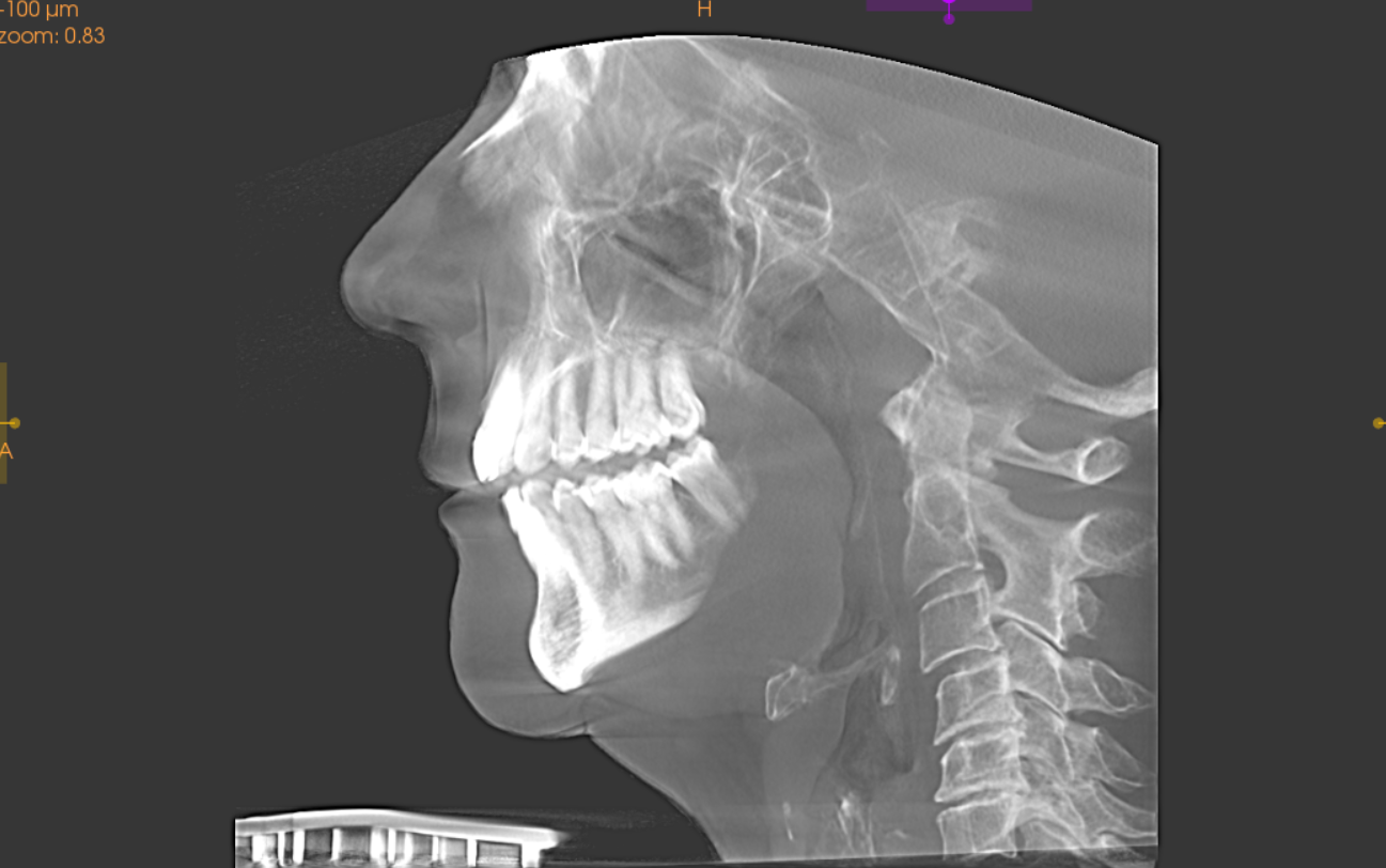
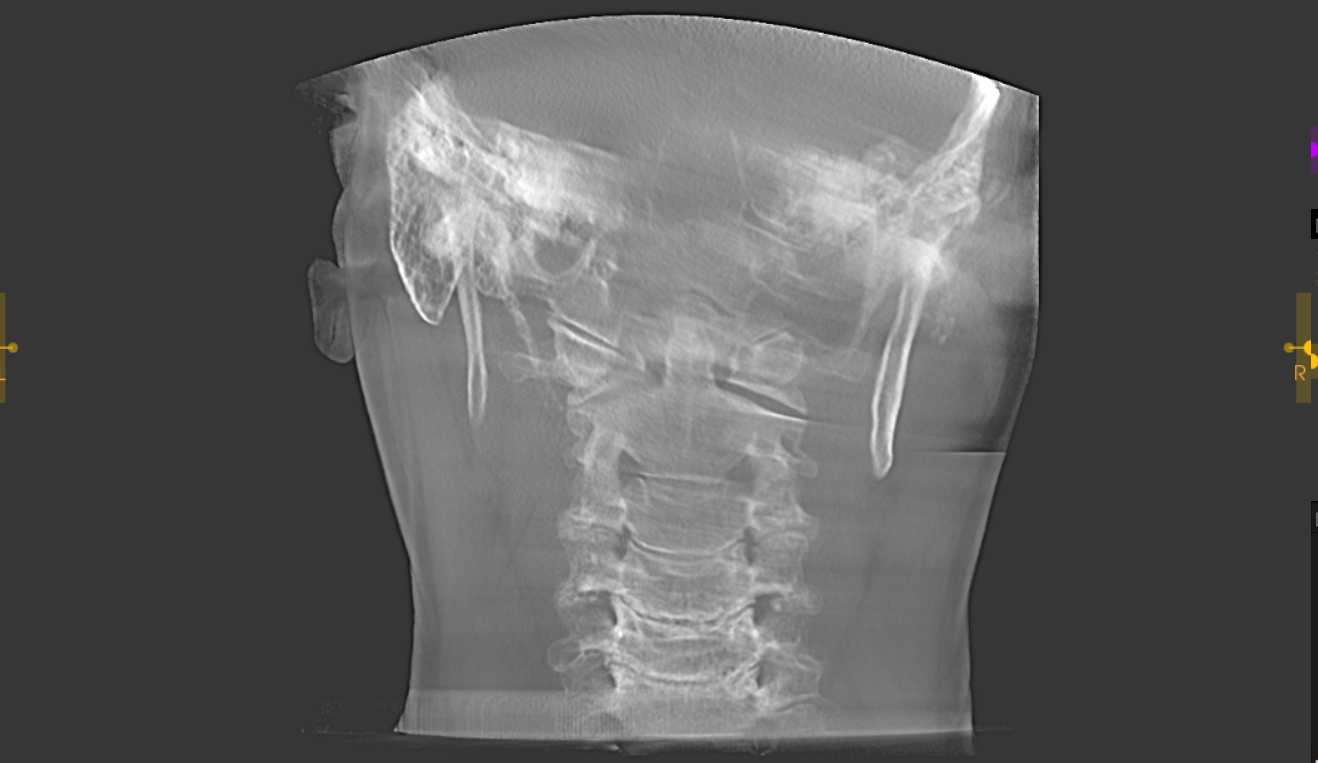
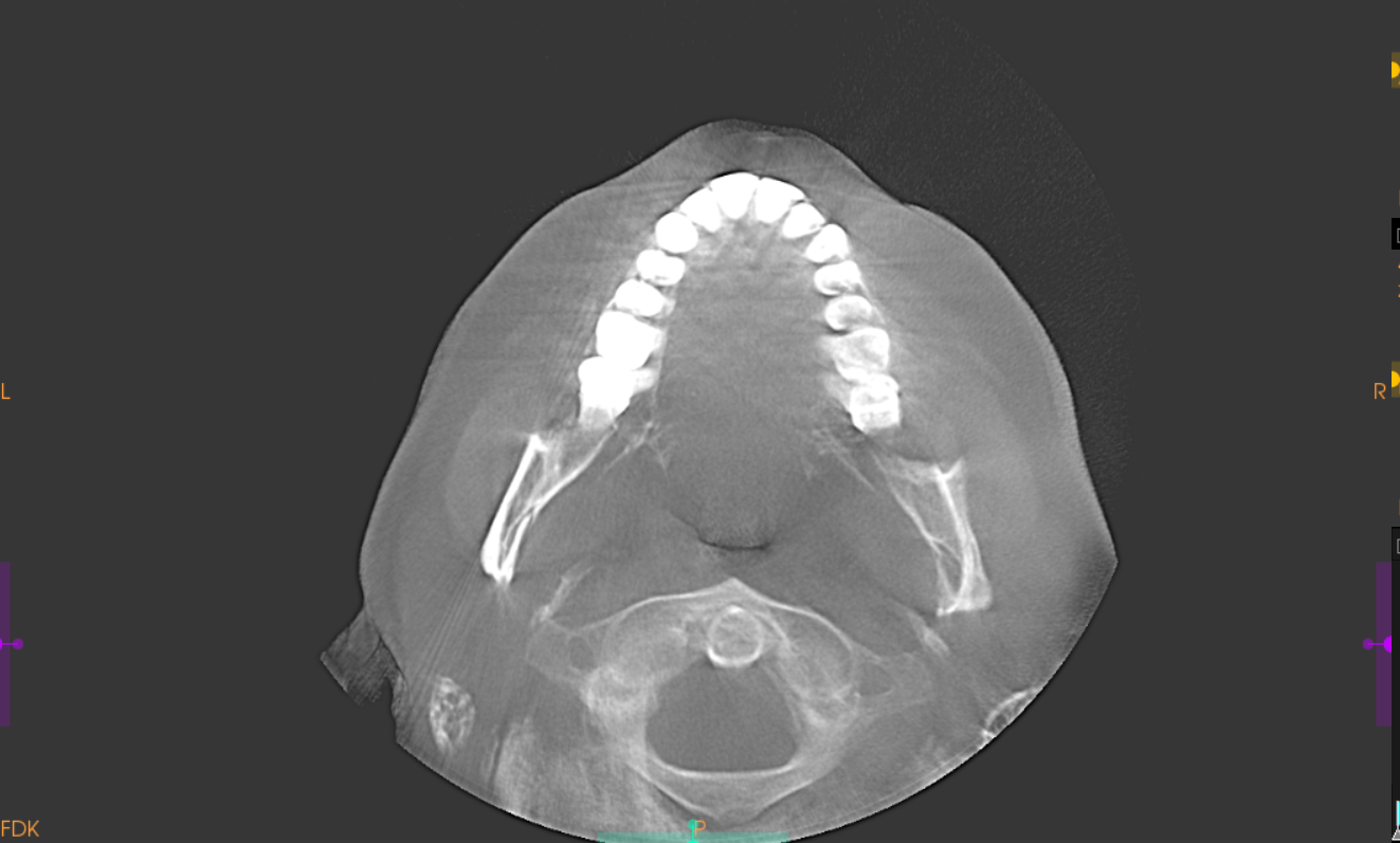
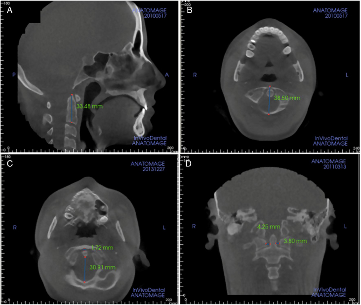
CBCT can provide high-resolution visualization of osseous anatomy with relatively efficient acquisition for appropriately selected indications. Its major strength at the CCJ is bone, not direct evaluation of the spinal cord, brainstem, ligaments, CSF or vascular structures.
Magnetic Resonance Imaging
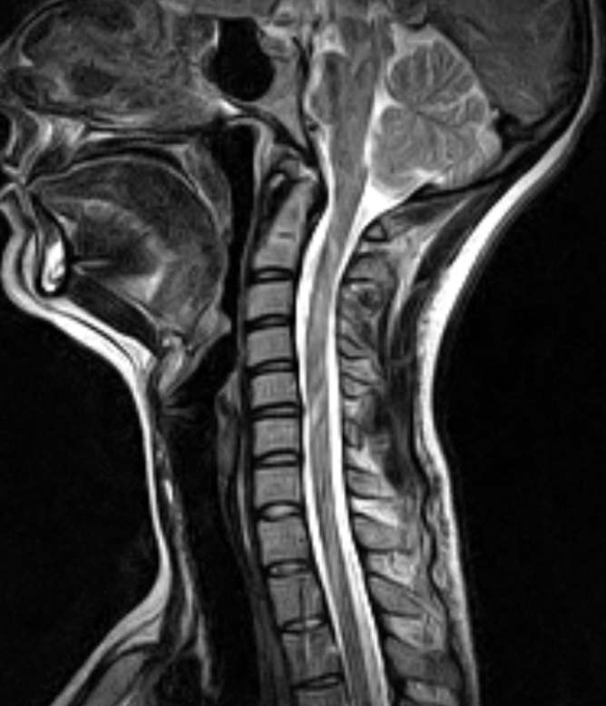
MRI is the most useful major modality for evaluating many of the soft tissues surrounding the CCJ. Depending upon protocol, it can assess:
- spinal cord
- brainstem
- cerebellar tonsils
- ligaments
- bone marrow
- neural compression
- soft-tissue injury
- syringomyelia
- CSF spaces
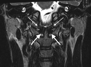
High-resolution MRI can demonstrate detailed morphology of the transverse and alar ligament complexes, although imaging appearance must always be interpreted in clinical context. 13
Abnormal MRI signal does not automatically equal mechanical instability.
That principle is particularly important when evaluating subtle ligament abnormalities after trauma.
Craniocervical Measurements
Several radiographic and MRI measurements have been developed to evaluate relationships between the skull and upper cervical spine. These include:
- Atlantodental interval (ADI)
- Basion-dens interval (BDI)
- Basion-axis interval (BAI)
- Powers ratio
- Clivo-axial angle (CXA)
- Grabb-Mapstone-Oakes / pB-C2 measurement
- McRae line
- Chamberlain line
- measurements of cerebellar tonsillar position
These measurements can provide valuable information, but they are not interchangeable and should not be interpreted in isolation.
Clivo-Axial Angle
The clivo-axial angle describes the angular relationship between the clivus and upper cervical spine. Published thresholds vary depending on methodology, patient population and imaging position. A systematic review focused on craniocervical instability in Ehlers-Danlos syndrome reported commonly used reference values around 145-160° and identified <135° as one threshold used for pathological kyphotic angulation in that literature. 14
That should not be converted into a universal rule that every person below a particular angle has brainstem compression.
Grabb-Oakes / pB-C2
The pB-C2 measurement estimates ventral encroachment upon the brainstem region. A value greater than approximately 9 mm has been used in specific clinical populations as an indicator of increased ventral brainstem encroachment. 14 Again, it is one measurement within a larger clinical and radiographic assessment. Age, imaging technique, head position and the clinical question affect how these measurements should be interpreted.
Trauma to the Craniocervical Junction
Serious CCJ injuries can follow substantial mechanical loading. Lower-energy trauma may also be significant in people with fragile bone or a pre-existing disorder. Potential mechanisms include:
- motor-vehicle collisions
- high-energy acceleration-deceleration
- axial loading
- falls from height
- diving injuries
- contact-sport trauma
- severe distraction
- combined flexion, extension and rotation
Traumatic injury can involve:
- occipital condyle fractures
- atlas fractures
- odontoid fractures
- transverse-ligament disruption
- alar-ligament injury
- atlantoaxial instability
- atlanto-occipital dissociation
- combined osteoligamentous injury
Because vital neural and vascular structures are nearby, severe CCJ injuries can be catastrophic. 1 A major principle is therefore:
A high-energy mechanism with concerning neurological or radiographic findings is not simply a mechanical neck problem.
It requires appropriate medical and imaging evaluation.
Symptoms Potentially Associated With the Upper Cervical Region
Disorders involving upper cervical structures may present with symptoms such as:
- upper neck pain
- suboccipital pain
- restricted cervical movement
- headache
- pain provoked by movement
- disequilibrium or dizziness in selected cervical conditions
- neurological symptoms in more serious pathology
However, none of these findings is specific for CCJ instability or ligament injury.
The Pediatric Craniocervical Junction
The developing pediatric CCJ deserves separate consideration. An infant’s upper cervical spine is not simply a miniature adult spine. Important differences include:
- proportionally larger head size
- incomplete ossification
- open synchondroses
- different facet geometry
- greater ligamentous laxity
- developing musculature
- greater overall mobility
- immature neuromuscular control
These differences affect both biomechanics and imaging interpretation. Normal developmental anatomy can sometimes resemble fracture or abnormality if adult criteria are applied to a young child. Published pediatric radiology literature therefore emphasizes knowledge of normal CCJ development when interpreting trauma or suspected pathology. 15
For a deeper discussion, see our guide to alar ligaments in babies and children.
When Findings Become More Concerning
Seek emergency care for sudden weakness, difficulty speaking or walking, a sudden severe unusual headache, or loss of consciousness, especially after trauma. These symptoms need urgent medical assessment.
Symptoms and imaging findings need to be interpreted together. Features warranting greater concern can include:
- substantial recent trauma
- progressive neurological deficit
- weakness or altered coordination
- myelopathic findings
- new cranial-nerve abnormalities
- severe or unusual occipital headache
- altered consciousness following trauma
- suspected fracture
- abnormal traumatic displacement
- suspected spinal-cord or brainstem compression
- suspected vascular injury
- other red flags suggesting pathology beyond uncomplicated mechanical neck pain
Appropriate referral and additional imaging is sometimes required along with chiropractic care of the upper cervical region.
Where Does Upper Cervical Chiropractic Fit?
Understanding CCJ anatomy is valuable in upper cervical chiropractic. A responsible upper cervical evaluation may incorporate:
- detailed history
- neurological examination
- orthopedic examination
- cervical range of motion
- sensorimotor and proprioceptive assessment when appropriate
- review of existing imaging
- obtaining or referring for additional imaging when clinically indicated
- identification of red flags and contraindications
- conservative management when the clinical presentation is appropriate
Additional manual therapy and exercise may have a role in selected neuromusculoskeletal neck-related presentations. Those conditions require diagnosis and management appropriate to the underlying pathology. The clinical value of understanding the CCJ is therefore not in attributing every neurological symptom to “C1 misalignment.” It is in recognizing how anatomy, biomechanics, neurological function, imaging and clinical examination interact and understanding when conservative care is appropriate and when it is not.
NeckCare at Apex Chiropractic
At Apex Chiropractic, we use NeckCare as part of the initial examination for patients other than babies and small children. It helps assess:
- Range of motion: how far the neck moves.
- Joint position error: how accurately a person returns their head to a target position.
- Sensorimotor control: how well head movement is coordinated during the assessment.
These findings complement the history and clinical examination. They do not independently diagnose a torn ligament, craniocervical instability or the cause of dizziness.
The Craniocervical Junction Is a System
Perhaps the most important concept is also the simplest:
The craniocervical junction cannot be understood by studying one bone, one ligament or one measurement in isolation.
Its stability depends upon the interaction of:
Bone
Occiput, atlas, axis and their articular geometry.
Ligaments
Including the transverse ligament, alar ligaments, tectorial membrane and capsular structures.
Muscles
Particularly the deep upper cervical and suboccipital musculature.
Neuromuscular control
Continuous proprioceptive feedback and sensorimotor regulation.
Neural anatomy
Brainstem, spinal cord and upper cervical afferent pathways.
Vascular anatomy
Including the highly mobile V3 segment of the vertebral arteries.
Meningeal and CSF anatomy
Including the spinal dura, foramen magnum, subarachnoid space and myodural connections.
The remarkable feature of the craniocervical junction is that it achieves substantial mobility while protecting some of the most important structures in the human body. Understanding that balance between mobility and stability is fundamental to interpreting CCJ anatomy, biomechanics, imaging and clinical findings correctly.
Putting the Findings in Context
The craniocervical junction is an integrated biomechanical and neurological system in which bone, ligaments, joints, muscles, sensory control, neural structures, vascular anatomy and cerebrospinal-fluid spaces function together.
A finding involving one component should therefore be interpreted within the context of the entire system and not in isolation.
Talk With Us About Your Neck Symptoms
If you have ongoing neck pain, restricted movement or headaches, we can discuss whether a cervical evaluation is appropriate and whether additional medical assessment is needed. If you are not local to our clinic, you can request a telehealth video conference to review your specific case and any imaging that you already have. Request an appointment or call (904) 310-4176.
References
- Offiah CE, Day E. The craniocervical junction: embryology, anatomy, biomechanics and imaging in blunt trauma. Insights Imaging. 2017;8:29–47.
- Lindenmann J, et al. Kinematics of the Cervical Spine Under Healthy and Degenerative Conditions: A Systematic Review. 2022.
- Fiester P, et al. Anatomic, functional, and radiographic review of the ligaments of the craniocervical junction. J Craniovertebr Junction Spine. 2021;12(1):4–9.
- Effects of occipital-atlas stabilization on upper cervical spine rotation combinations: an in vitro study. 2023.
- Peng B, et al. Cervical Proprioception Impairment in Neck Pain—Pathophysiology, Clinical Evaluation, and Management: A Narrative Review. Pain Ther. 2021.
- Bogduk N. The anatomical basis for cervicogenic headache. J Manipulative Physiol Ther. 1992;15(1):67–70.
- Zhang L, et al. The myodural bridge complex: a comprehensive review of morphology, physiology, developmental biology and pathology. Front Med. 2026. doi:10.3389/fmed.2026.1790220.
- Enix DE, Scali F, Pontell ME. The cervical myodural bridge, a review of literature and clinical implications. J Can Chiropr Assoc. 2014;58(2):184–192.
- Prevalence of anatomical variations at the suboccipital (V3) segment of the vertebral artery: a systematic review. 2023.
- Korbecki A, et al. Imaging of cerebrospinal fluid flow: fundamentals, techniques, and clinical applications of phase-contrast magnetic resonance imaging. Pol J Radiol. 2019.
- Characterization of Cyclic CSF Flow in the Foramen Magnum and Upper Cervical Spinal Canal with MR Flow Imaging and Computational Fluid Dynamics. 2010.
- American College of Radiology. ACR Appropriateness Criteria: Acute Spinal Trauma.
- Magnetic resonance imaging anatomy of the craniovertebral ligaments: A radiological study with confirmatory dissection. J Craniovertebr Junction Spine. 2022;13(3):309–317.
- Lohkamp LN, Marathe N, Fehlings MG. Craniocervical Instability in Ehlers-Danlos Syndrome—A Systematic Review of Diagnostic and Surgical Treatment Criteria. Global Spine J. 2022.
- Sriram R, et al. CT evaluation of pediatric craniocervical distraction injuries. 2026.
- Carrasco-Uribarren A, et al. Is manual therapy effective for cervical dizziness? A systematic review and meta-analysis of randomized controlled trials. BMC Musculoskelet Disord. 2025;26:659.
- Chacko N, et al. Manual therapy with exercise for neck pain. Cochrane Database Syst Rev. 2025;12:CD011225.
- Spinal manipulation for the management of cervicogenic headache: a systematic review and meta-analysis. 2020.