By William Soriano, DC | Upper Cervical Chiropractor | Apex Chiropractic
The alar ligaments are paired stabilizing ligaments of the craniocervical junction, the place where the neck and head meet. These ligaments extend primarily from the lateral aspects of the odontoid process of the axis(C2), toward the medial surfaces of the occipital condyles(C0).
Their principal mechanical role is to restrain/check excessive axial rotation, lateral bending, and, to a lesser degree, flexion-extension motion of the head and upper cervical spine. They function bilaterally as part of an integrated stabilizing system rather than standalone stabilizers.
Alar ligament injury may happen when craniocervical trauma induces severe rotational, lateral-bending, distraction, or high-energy forces. However, diagnosing an isolated alar ligament injury/tear is difficult. Symptoms can be nonspecific and physical examination cannot independently establish the injury/tear. Plus, conventional MRI findings may be variable or difficult to interpret, especially if the right slice, stack, or sequences are not properly ordered/taken.
An alar ligament injury cannot be determined by any one single method. Instead, take into account and integrate:
- Mechanism of injury
- Neurological findings
- Craniocervical alignment
- Osseous imaging
- Ligament-sensitive MRI when indicated
- Dynamic imaging in selected cases
- Specialist consultation when instability is suspected
An abnormal signal within an alar ligament does not automatically mean a traumatic rupture or clinically meaningful instability is present.
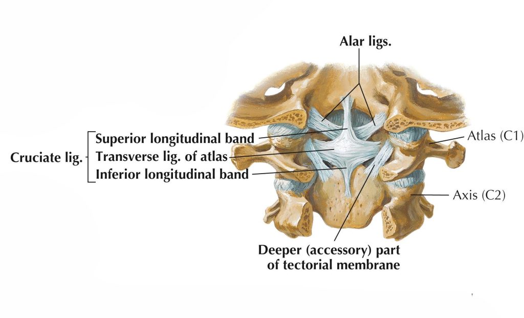
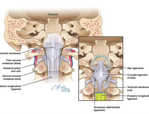
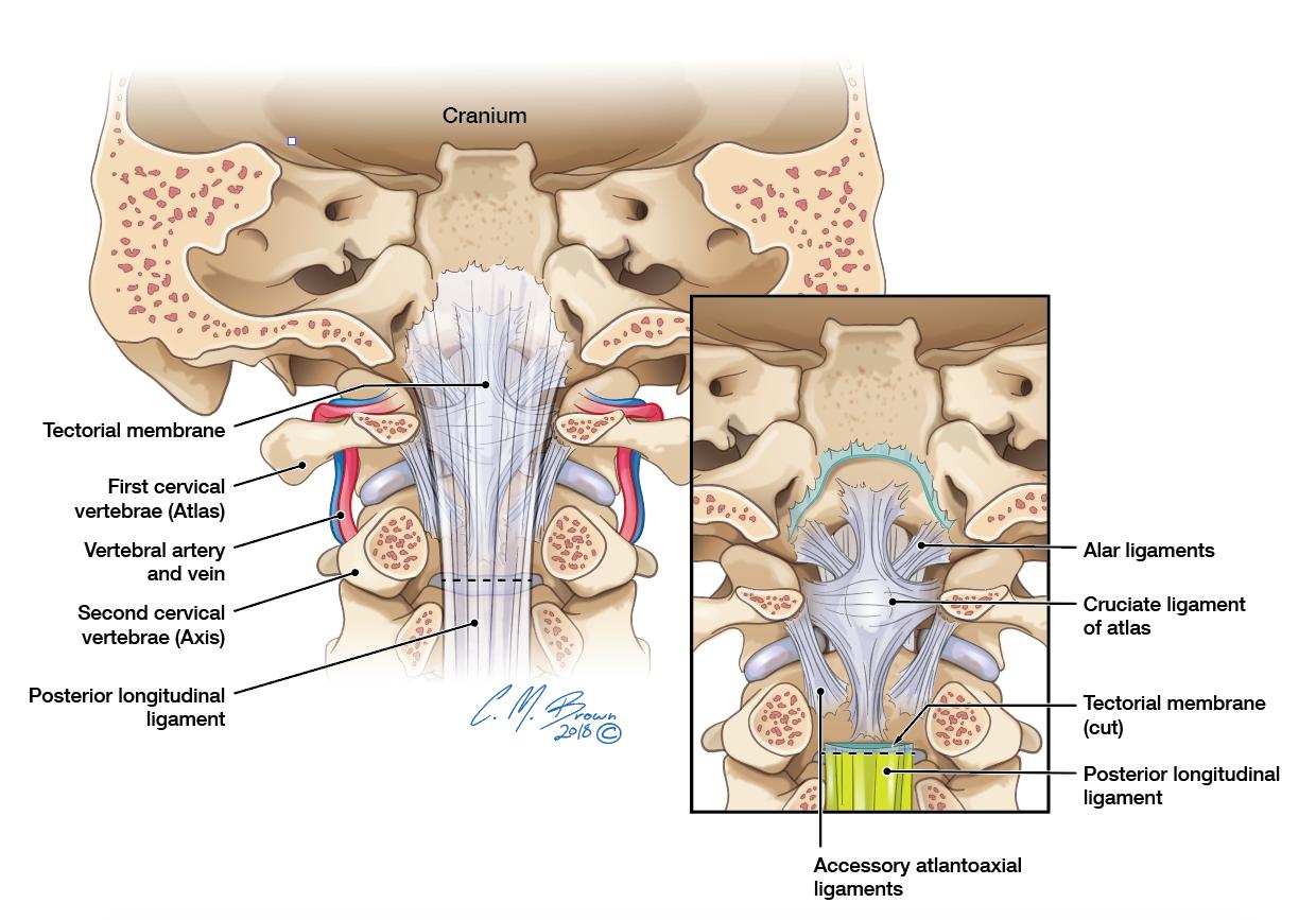
Figure 1. Overview of the craniocervical junction demonstrating the occiput, atlas (C1), axis (C2), odontoid process, and major stabilizing ligaments.
The alar ligaments are two short, strong bands of dense connective tissue located at the craniocervical junction. Alar derives from the Latin word for “wing.” When viewed from the front, the right and left ligaments extend outward and upward from the odontoid region in a wing-like fashion.
They form part of the ligamentous complex connecting:
- The occiput
- The atlas
- The axis
- The odontoid process
Their main purpose is to regulate and limit excessive motion between the skull and the upper cervical spine. The alar ligaments are located deep within the craniocervical junction.
Located:
- Lateral to the apical ligament of the dens
- Anterior to portions of the tectorial membrane
- Superior and lateral to the transverse ligament
- Between the dens and the medial occipital condylar region
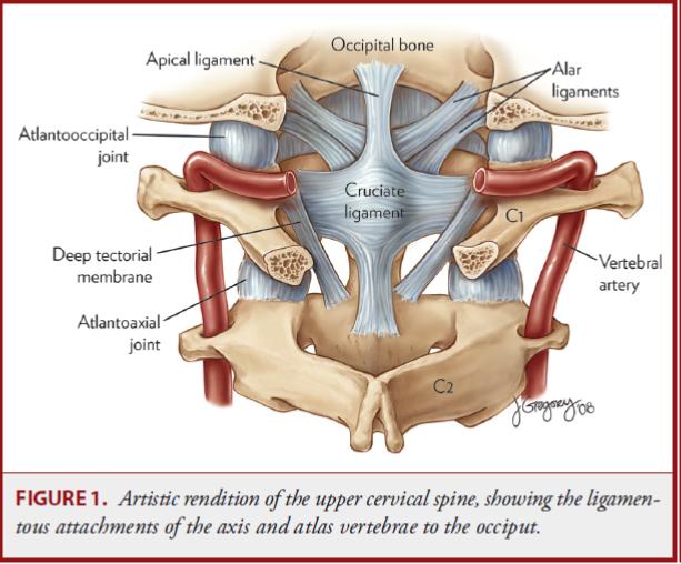
Figure 2. The paired alar ligaments extend from the lateral aspects of the dens to the medial occipital condyles.
The ligaments are typically not visible on standard X-rays or CBCT because these modalities primarily depict mineralized tissue. MRI is the principal imaging used when direct evaluation of ligament integrity is indicated. Each alar ligament typically originates from the upper lateral surface of the odontoid process.
The exact attachment may vary in:
- Height
- Width
- Fiber orientation
- Shape
- Relationship to adjacent connective tissues
The dens attachment is generally narrower than the occipital attachment.The fibers then travel superiorly and laterally to attach primarily to the medial surfaces of the occipital condyles.This orientation allows the ligaments to span the transition between the axis and skull while crossing both the atlantoaxial(C1/C2) and atlanto-occipital(C0/C1) regions.
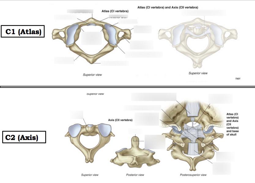
Figure 3. Atlas and axis demonstrating the osseous relationships supporting the alar ligaments.
Some anatomical descriptions may include fibers extending toward the lateral mass or other components of the atlas, but the presence of a distinct atlantal portion has not been consistently established.This variability is important because simplified anatomical diagrams may imply that every person has identical, sharply defined alar ligament bundles. Actual anatomy is more complex, especially in this area.
The alar ligament fibers and shape are described as:
- Cord-like
- Fan-shaped
- Ovoid
- Rounded
- V-shaped when viewed together
Fiber orientation varies among individuals. Fibers may travel:
- Nearly horizontally
- Superolaterally
- Slightly inferolaterally
- In mixed bundles with different orientations
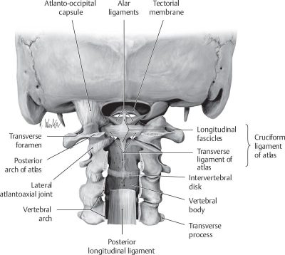
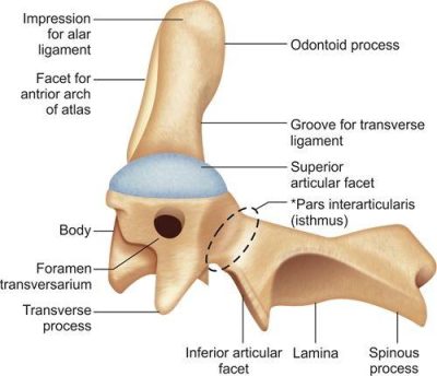
Figures 4/5. Posterior view illustrating the relationship between the occipital condyles, atlas, axis, and alar ligaments and odontoid process demonstrating the attachment site of the alar ligaments.
Orientation influences when each ligament becomes tensioned during movement. A more horizontal ligament may respond differently to rotation than a ligament with a stronger superior or inferior orientation. Therefore, anatomical variation may influence both normal motion and how ligaments appear on MRI.
The alar ligaments are composed primarily of collagen-rich dense connective tissue organized to resist tensile loading forces. In a cadaveric tensile study involving 19 ligament specimens, failure occurred between 87 and 346 newtons, with a mean failure force of approximately 187 newtons. However, the specimens came from older adult cadavers, meaning these values should not be treated as universal thresholds for living patients of all ages. Also, laboratory failure does not represent the exact force necessary to produce injury during a real world collision. Biological injury largely depends on:
- Direction of force
- Speed of loading
- Head position
- Repetition
- Combined rotation and translation
- Bone quality
- Age
- Existing degeneration
- Congenital anatomy
- Contributions from surrounding ligaments
The alar ligaments are particularly important in restraining excessive upper cervical axial rotation. During rotation of the head, the ligament opposite the direction of rotation will become more tensioned.
For example:
- Right head rotation tensions the left alar ligament.
- Left head rotation tensions the right alar ligament.
This description is useful, but it should not be interpreted as an absolute on-off mechanism. Both ligaments interact with the transverse ligament, facet joints, joint capsules, tectorial membrane, muscles, and osseous geometry. Cadaveric studies have demonstrated increased motion at both C0-C1 and C1-C2 following alar ligament transection. More recent robotic testing found that unilateral alar ligament sectioning increased lateral bending, axial rotation, and flexion-extension motion.
The same study estimated that the intact alar ligaments contributed approximately 19% of resistance to axial rotation and 16% of resistance to lateral bending under the tested conditions.These figures describe one experimental model and should not be interpreted as fixed percentages in every living patient.
Lateral Bending
The alar ligaments also limit excessive lateral bending. Lateral bending produces coupled motion within the craniocervical junction. Because the ligaments cross from the dens to the occiput, side bending changes their length and tension.The opposite-side ligament is generally expected to become more tensioned, although fiber orientation and coupled rotation may alter the precise loading pattern.
Flexion and Extension
The alar ligaments are not the primary restraints to sagittal-plane motion, but experimental evidence shows that they contribute to stability in both flexion and extension.Their influence should therefore be understood as multidirectional rather than limited exclusively to rotation.The ligaments work as a paired system.
Mechanical modeling suggests that injury to one ligament may alter how the entire bilateral restraint mechanism functions. The remaining ligament does not necessarily act as a complete substitute for the injured side.
Relationship to Other Craniocervical Stabilizers
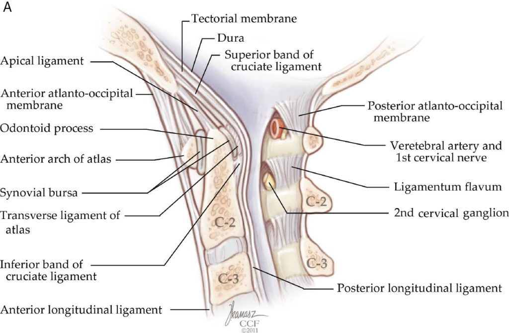
Figure 6. The major stabilizing ligaments of the craniocervical junction.
The alar ligaments should never be evaluated in isolation. Craniocervical stability depends on the combined function of:
- Transverse ligament of the atlas
- Cruciform ligament
- Tectorial membrane
- Atlanto-occipital joint capsules
- Atlantoaxial joint capsules
- Apical ligament
- Anterior and posterior membranes
- Osseous congruity
- Suboccipital musculature
- Neuromuscular control
The transverse ligament is especially important in restraining anterior translation of the atlas relative to the dens.The alar ligaments primarily regulate rotational and lateral motion, but they also assist in preventing excessive displacement when the broader stabilizing system is stressed.
A patient may therefore have:
- An isolated alar abnormality
- A combined alar and capsular injury
- Multiligamentous injury
- Osseous injury with secondary ligament compromise
- MRI signal changes without mechanical instability
These are not equivalent clinical situations.
Injury Mechanisms
Alar ligament injury is most plausible when the head and upper cervical spine are exposed to substantial:
- Axial rotation
- Lateral bending
- Distraction
- Flexion-extension
- Combined multiplanar loading
Potential mechanisms include:
- Motor-vehicle collision
- High-energy whiplash
- Contact-sport collision
- Diving injury
- Fall from height
- Direct cranial impact
- Craniocervical distraction
- Severe rotational trauma
Minor neck pain alone is insufficient evidence of alar ligament injury, however, the probability of meaningful ligament disruption increases when the mechanism is accompanied by:
- Fracture
- Dislocation
- Abnormal craniocervical alignment
- Neurological deficits
- Severe occipital pain
- Marked guarding
- Altered consciousness
- Other indicators of high-energy trauma
Possible Clinical Presentation
There is no symptom pattern that is specific enough to diagnose an alar ligament injury without further evaluation. The following presentations may raise suspicion of alar ligament involvement.
Potential findings may include:
- Upper cervical or suboccipital pain
- Pain aggravated by rotation
- Restricted or guarded head movement
- Sensation of instability
- Headache
- Dizziness
- Visual-motion sensitivity
- Nausea
- Disequilibrium
- Neurological complaints after trauma
These symptoms overlap with many other disorders, including:
- Cervical muscle injury
- Facet-joint injury
- Concussion
- Vestibular disorders
- Migraine
- Occipital neuralgia
- Vertebral artery pathology
- Upper cervical fracture
- Atlantoaxial instability
- Craniocervical dissociation
Symptoms alone cannot determine ligament integrity.
Physical Examination
Alar Ligament Stress Tests
Clinical tests described for evaluating possible alar ligament compromise include:
- Rotation stress test
- Side-bending stress test
- Lateral shear-type testing
- C2 spinous-process or “kick” test
- Bending-rotation test
The theoretical basis is that controlled movement of the skull should produce early tension through the alar ligament and associated motion of C2.
Interpretation Limitations
Manual examination has important limitations.
A positive test may reflect:
- Pain
- Muscle guarding
- Normal anatomical variation
- Capsular restriction
- Examiner technique
- Generalized hypermobility
- Apprehension
Cadaveric biomechanical testing demonstrated measurable motion changes after alar ligament transection, but the investigators specifically noted that it remains uncertain whether clinicians can reliably feel changes of that magnitude. A small diagnostic study reported moderate accuracy for several manual alar ligament tests when compared with MRI, but the sample was limited and MRI itself is not a perfect gold standard for functional instability. Clinical testing should therefore be regarded as part of a screening process, not definitive proof of injury.
Safety Principle
Provocative upper cervical stress testing should not be performed aggressively when the patient has:
- Recent high-energy trauma
- Suspected fracture
- Suspected dislocation
- Severe neurological symptoms
- Signs of craniocervical instability
- Possible vascular emergency
- Marked midline tenderness
- Inability to actively control the head
In these situations, stabilization and appropriate medical imaging take priority.
Imaging Radiographs
Routine radiographs cannot directly visualize the alar ligaments, but they may demonstrate indirect evidence of instability, including:
- Abnormal alignment
- Lateral-mass asymmetry
- Increased atlantodental interval
- Fracture
- Translation
- Abnormal motion on appropriately indicated dynamic studies
Apparent asymmetry on open-mouth imaging may also result from head rotation, positioning, beam geometry, or normal anatomy.
CT and CBCT
CT and CBCT provide excellent visualization of:
- Occipital condyles
- Atlas
- Axis
- Odontoid process
- Joint alignment
- Fractures
- Congenital variants
- Degenerative change
However, a structurally intact-looking bone study does not directly establish normal alar ligament tissue. CT is generally favored in acute trauma because it is fast and highly effective for identifying fracture and gross osseous malalignment. CBCT can provide detailed osseous anatomy at relatively high spatial resolution, but it is not a substitute for MRI when direct ligament or neural-tissue evaluation is required.
MRI
MRI may visualize the alar ligaments using high-resolution, thin-section sequences oriented to the craniocervical junction.
Potential MRI observations include:
- Fiber continuity
- Ligament thickness
- Signal intensity
- Asymmetry
- Surrounding edema
- Associated tectorial or transverse-ligament injury
- Hemorrhage
- Joint effusion
- Neural or soft-tissue injury
MRI interpretation is technically difficult because the ligaments are small, obliquely oriented, and anatomically variable.
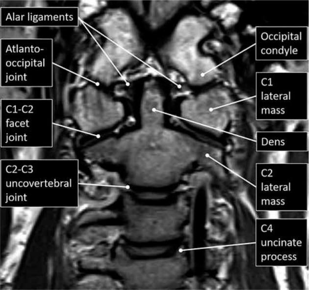
Figure 7. Normal coronal MRI of the craniocervical junction.
MRI Signal Changes and Whiplash
Earlier studies reported increased high-signal changes in the alar ligaments of some patients with chronic whiplash symptoms. These observations led to proposals that MRI could identify persistent ligament injury. Subsequent research raised substantial concerns about this interpretation.
High-signal changes have also been identified in:
- Uninjured individuals
- Patients with nontraumatic neck pain
- People without demonstrable instability
Prospective research found that upper cervical ligament high-signal changes after lower-grade whiplash were not predictive of outcome at 12 months. Follow-up imaging also found that these signals did not meaningfully change over the first year and were similarly prevalent in whiplash patients and noninjured patients with chronic neck pain. The investigators concluded that some of these appearances were more likely normal variants than traumatic lesions. Therefore, high signal within an alar ligament is not, by itself, proof of traumatic rupture, instability, or the cause of a patient’s symptoms.
Reliability of MRI Diagnosis
Even in major craniocervical trauma, MRI assessment of the alar ligaments can show inconsistent agreement between observers. One study evaluating MRI for ligament injury in atlanto-occipital dissociation found unsatisfactory interobserver and intraobserver reliability for alar ligament assessment. The authors emphasized the need for standardized imaging and interpretation protocols.This does not mean MRI is useless, it means MRI findings must be interpreted alongside:
- Trauma mechanism
- CT findings
- Neurological status
- Alignment measurements
- Other ligament findings
- Dynamic stability when appropriate
- Clinical progression
Isolated Alar Ligament Injury
Isolated alar ligament injury appears to be uncommon and is reported primarily through small case series and case reports.The available literature does not establish one universally accepted treatment protocol. Management has included:
- Temporary rigid immobilization
- Activity restriction
- Clinical observation
- Repeat imaging
- Dynamic CT or radiography
- Specialist consultation
- Gradual return to activity after stability is established
A 2024 case review emphasized that the significance of treating an MRI finding alone remains uncertain and that clinical recovery and demonstration of stability are important components of decision-making.
Differential Diagnosis
When alar ligament injury is considered, the differential diagnosis should include:
Osseous Injury
- Occipital-condyle fracture
- Atlas fracture
- Odontoid fracture
- Axis fracture
- Atlanto-occipital dislocation
- Atlantoaxial dislocation
Ligamentous Injury
- Transverse-ligament injury
- Tectorial-membrane injury
- Capsular injury
- Multiligamentous craniocervical injury
Neurological and Vascular Conditions
- Spinal cord injury
- Brainstem injury
- Vertebral artery dissection
- Concussion
- Traumatic brain injury
- Cervical nerve injury
Musculoskeletal Conditions
- Suboccipital strain
- Facet-joint injury
- Cervicogenic headache
- Occipital neuralgia
- Myofascial pain
- Temporomandibular dysfunction
Vestibular Conditions
- BPPV
- Peripheral vestibular injury
- Vestibular migraine
- Persistent postural-perceptual dizziness
Red Flags Requiring Urgent Medical Evaluation
Urgent assessment is warranted after trauma when the patient presents with:
- New limb weakness
- New numbness or loss of coordination
- Difficulty walking
- Loss of bladder or bowel control
- Altered consciousness
- Severe or rapidly worsening occipital pain
- Cranial-nerve abnormalities
- Difficulty swallowing or speaking
- Double vision
- Drop attacks
- New facial sensory changes
- Inability to support or control the head
- Signs of fracture or dislocation
- Symptoms concerning for vascular injury
These findings should not be attributed to a routine upper cervical “misalignment” without appropriate emergency evaluation.
Upper Cervical Chiropractic Considerations
The alar ligaments are directly relevant to upper cervical practice because they regulate motion between the skull, atlas, and axis. However, their clinical significance must be presented accurately.
What Can Be Stated Confidently
- The alar ligaments contribute to upper cervical rotational and lateral stability.
- Trauma can injure the alar ligaments.
- Loss of ligament integrity may increase craniocervical motion.
- CBCT cannot directly establish ligament integrity.
- MRI findings require clinical correlation.
- Suspected instability changes the risk profile of manual treatment.
What Should Not Be Claimed Without Evidence
- That atlas displacement automatically means alar ligament injury
- That a rotated atlas on static imaging proves ligament laxity
- That MRI signal automatically represents traumatic tearing
- That dizziness confirms alar ligament pathology
- That manual palpation can definitively diagnose a tear
- That an upper cervical adjustment repairs a structurally torn ligament
- That every patient with whiplash requires specialized alar ligament MRI
Treatment-Safety Principle
When significant ligamentous instability is suspected, forceful end-range rotation, aggressive stress testing, and high-velocity procedures should be avoided until instability has been appropriately investigated.
Upper cervical care should be considered safe and appropriate after:
- Fracture has been excluded
- Major instability has been excluded
- Neurological status is understood
- Imaging has been reviewed where clinically indicated
- The selected procedure is compatible with the patient’s structural condition
Dizziness and Neurological Symptoms
The upper cervical spine contributes sensory input to systems involved in head-position awareness, balance, eye-head coordination, and postural control. An injury affecting the upper cervical joints, muscles, or ligaments could plausibly alter afferent input from the region. However, a direct chain such as alar ligament abnormality to brainstem dysfunction to then dizziness cannot be assumed in an individual patient without supporting evidence.
Dizziness after trauma may instead arise from:
- Concussion
- BPPV
- Vestibular injury
- Migraine
- Medication effects
- Autonomic dysfunction
- Cervical sensorimotor disturbance
- Psychological and perceptual factors
- Multiple simultaneous causes
The alar ligaments are therefore clinically relevant to upper cervical stability but should not be used as a universal explanation for post-traumatic neurological symptoms.
Patient Explanation
The alar ligaments are two small but important stabilizing ligaments near the top of the neck. They connect the second cervical vertebra to the base of the skull and help prevent the head from rotating or bending too far. A major accident can potentially stretch or tear these ligaments, but the symptoms are not unique. Neck pain, headache, dizziness, and stiffness can result from many different injuries.
The ligaments are also difficult to evaluate. They cannot be seen directly on standard X-rays or CBCT, and even MRI findings may not always distinguish injury from normal anatomical variation. For this reason, diagnosis should be based on the entire clinical picture rather than one symptom, one physical test, or one imaging observation.
FAQ
Can an alar ligament heal?
A partial ligament injury may heal or scar over time, but healing depends on the severity of injury, blood supply, mechanical stability, age, health status, and whether the region is repeatedly stressed. Complete disruption associated with instability requires specialist management.
Can CBCT show an alar ligament tear?
No. CBCT may show fractures, alignment, joint relationships, and congenital anatomy, but it does not directly demonstrate the soft-tissue fibers of the alar ligaments.
Is MRI definitive?
No. MRI is the most useful modality for direct ligament assessment, but image quality, sequence selection, anatomy, reader experience, and normal variation affect interpretation.
Can a patient have abnormal MRI signal without instability?
Yes. High-signal changes have been reported in people without acute injury and do not necessarily represent traumatic rupture or mechanical instability.
Can the ligaments cause dizziness?
Upper cervical injury may contribute to altered sensorimotor input, but dizziness has many possible causes. An alar ligament abnormality should not be assumed to be the cause without a broader neurological, vestibular, vascular, and cervical evaluation.
Can an adjustment repair a torn ligament?
A manual procedure does not biologically reconnect a completely torn ligament. Management depends on the severity of injury and stability of the craniocervical junction.
Should the alar ligaments be tested after every whiplash injury?
Not necessarily. Testing should be guided by injury mechanism, symptoms, examination findings, neurological status, and clinical suspicion. Aggressive testing may be inappropriate in acute or high-risk trauma.
References
- Dvorak J, Panjabi MM. Functional anatomy of the alar ligaments. Spine (Phila Pa 1976). 1987;12(2):183–189. doi:10.1097/00007632-198703000-00016. PMID: 3589810.
- Dvorak J, Schneider E, Saldinger P, Rahn B. Biomechanics of the craniocervical region: the alar and transverse ligaments. J Orthop Res. 1988;6(3):452–461. doi:10.1002/jor.1100060317. PMID: 3357093.
- Osmotherly PG, Rivett DA, Rowe LJ. Revisiting the clinical anatomy of the alar ligaments. Eur Spine J. 2013;22(1):60–64. doi:10.1007/s00586-012-2493-4. PMID: 22968541. PMCID: PMC3540300.
- Iwanaga J, Sardi JP, Laws T, Chapman JR, Oskouian RJ, Tubbs RS. Anatomy of Alar Ligament Part III: Biomechanical Study. World Neurosurg. 2017;107:1012–1015. doi:10.1016/j.wneu.2017.07.188.
- Tisherman R, Hartman R, Hariharan K, Vaudreuil N, Sowa G, Schneider M, Timko M, Bell K. Biomechanical contribution of the alar ligaments to upper cervical stability. J Biomech. 2020;99:109508. doi:10.1016/j.jbiomech.2019.109508. PMID: 31813563.
- von Piekartz H, Maloul R, Hoffmann M, Hall T, Ruch MM, Ballenberger N. Diagnostic accuracy and validity of three manual examination tests to identify alar ligament lesions: results of a blinded case-control study. J Man Manip Ther. 2019;27(2):83–91. doi:10.1080/10669817.2018.1539434. PMID: 30935337.
- Vetti N, Kråkenes J, Eide GE, Rørvik J, Gilhus NE, Espeland A. Are MRI high-signal changes of alar and transverse ligaments in acute whiplash injury related to outcome? BMC Musculoskelet Disord. 2010;11:260. doi:10.1186/1471-2474-11-260. PMID: 21070654. PMCID: PMC2989946.
- Vetti N, Kråkenes J, Ask T, et al. Follow-Up MR Imaging of the Alar and Transverse Ligaments after Whiplash Injury: A Prospective Controlled Study. AJNR Am J Neuroradiol. 2011;32(10):1836–1841. doi:10.3174/ajnr.A2636.
- Dyas AR, Niemeier TE, McGwin G, Theiss SM. Ability of magnetic resonance imaging to accurately determine alar ligament integrity in patients with atlanto-occipital injuries. J Craniovertebr Junction Spine. 2018;9(4):241–245. doi:10.4103/jcvjs.JCVJS_81_18. PMID: 30783347. PMCID: PMC6364359.
- Reeves BC, Valcarce-Aspegren M, Robert SM, et al. Isolated unilateral alar ligamentous injury: illustrative cases. J Neurosurg Case Lessons. 2024;7(14):CASE23664. doi:10.3171/CASE23664.