A guide to cerebellar tonsillar ectopia by William Soriano, DC | Upper Cervical Chiropractor | Apex Chiropractic
If your MRI report mentions “cerebellar tonsillar ectopia” or “low lying cerebellar tonsils,” you may wonder what that means. Could it explain your headaches, dizziness, neck pain, or balance problems? Does it mean you have Chiari malformation? Will you need surgery?
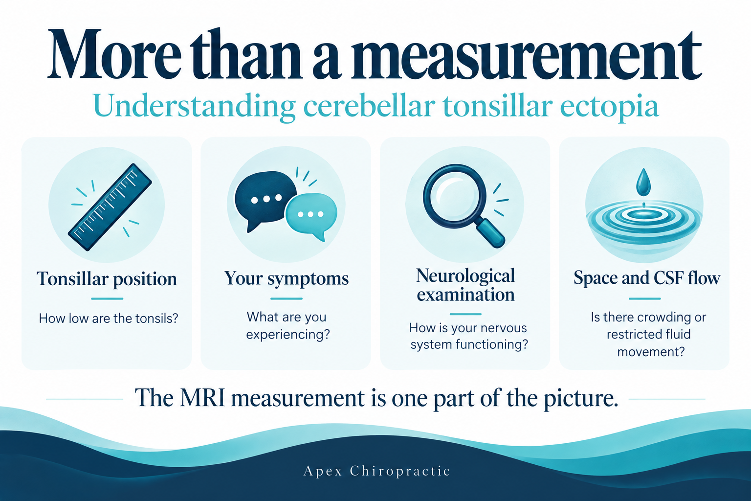
This finding does not automatically mean something is wrong or that treatment is necessary. Its importance depends on your symptoms, neurological examination, and the space around the brainstem and spinal cord. The movement of cerebrospinal fluid also matters. Understanding the complete picture is more useful than focusing on a single measurement.
What Are the Cerebellar Tonsils?
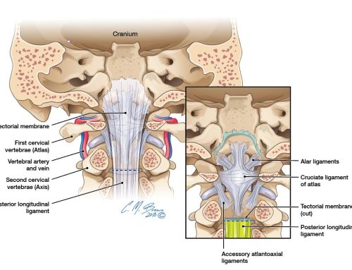
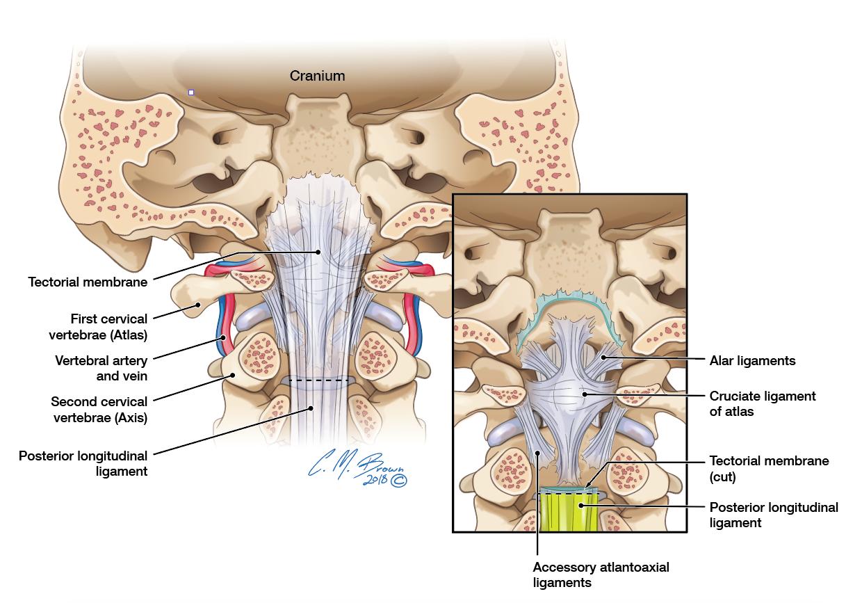
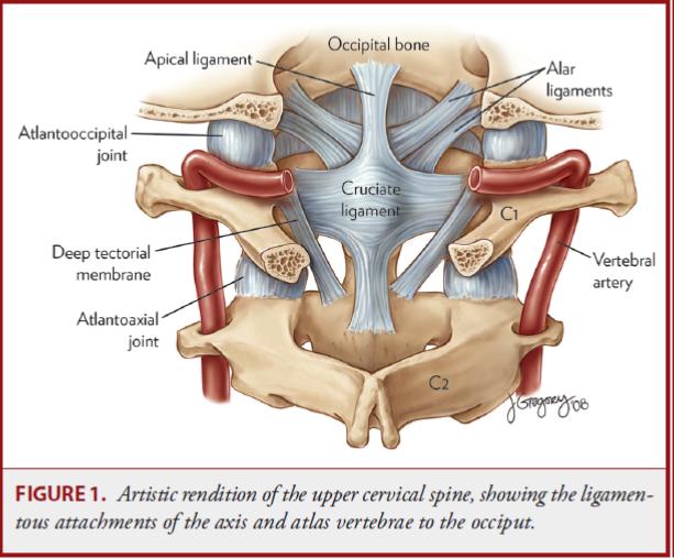
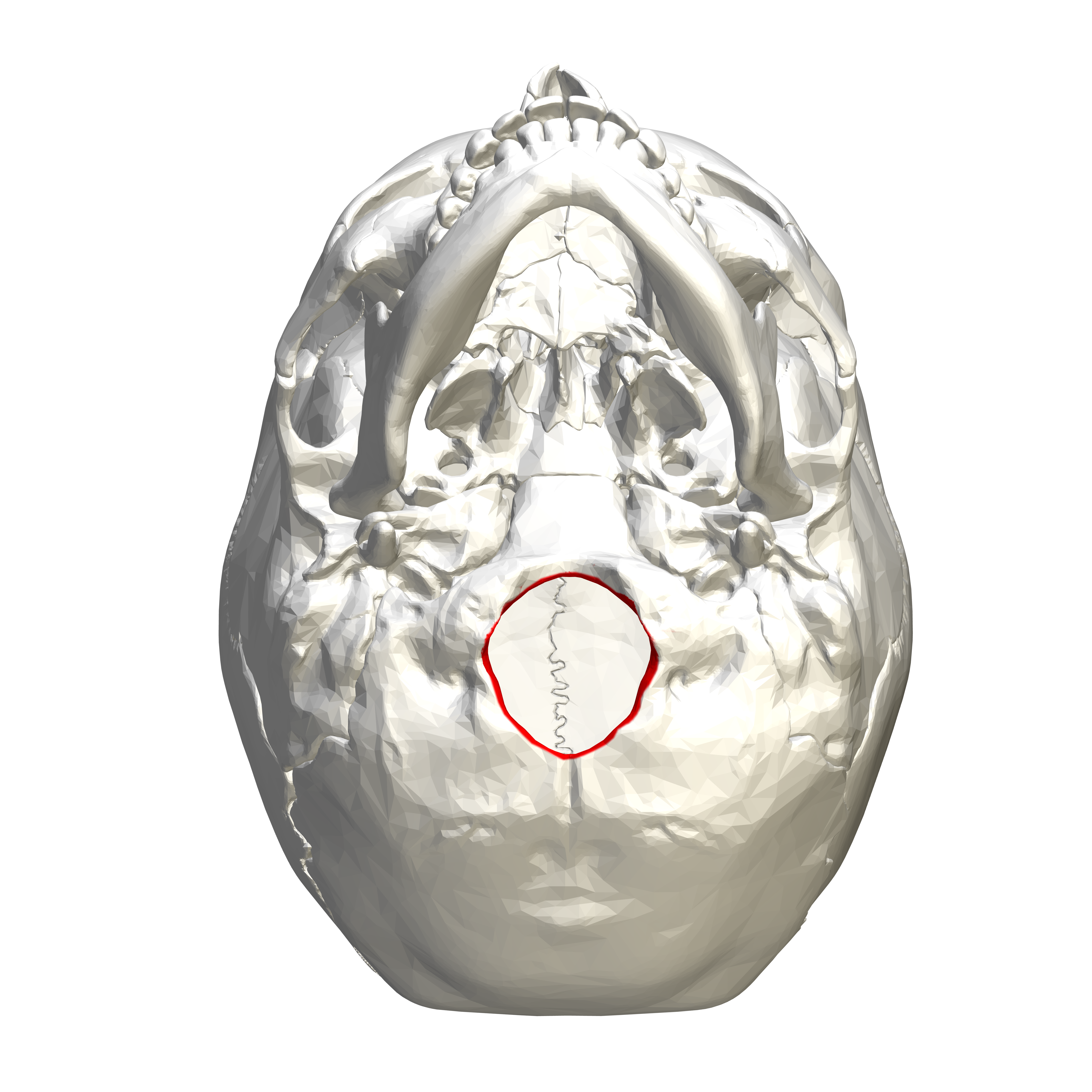
The cerebellum is the part of the brain that helps coordinate movement and balance. The cerebellar tonsils are two small, rounded areas on its underside. They are unrelated to the tonsils in your throat. Just below them is the foramen magnum, the large opening at the bottom of the skull. This is where the brainstem transitions into the spinal cord. The skull and the first two neck bones form the craniocervical junction. This region contains important nerves, blood vessels, supporting tissues, and spaces through which cerebrospinal fluid moves.
What Does Cerebellar Tonsillar Ectopia Mean?
Cerebellar tonsillar ectopia means that one or both cerebellar tonsils sit lower than expected, extending below the opening at the base of the skull.
An MRI report may call this:
- Low lying cerebellar tonsils
- Mild tonsillar descent
- Cerebellar tonsillar descent
These terms describe anatomy. They do not, by themselves, explain why someone has symptoms. Some people have this finding and feel completely well. Others have crowding or related abnormalities that deserve further evaluation.
Cerebellar Tonsillar Ectopia and Chiari Malformation
Not always.
Chiari I malformation is commonly associated with cerebellar tonsils extending approximately 5 millimeters or more below the foramen magnum. However, the measurement alone does not tell the whole story. A smaller amount of descent can sometimes occur with meaningful crowding. A larger amount may be present without significant symptoms. The important question is whether there is enough room for the nearby neurological structures and normal fluid movement. Clinicians also consider the shape of the tonsils, symptoms, examination findings, and associated abnormalities. A diagnosis should bring these pieces together rather than depend on a number alone.[1]
Why CSF Flow Matters in Cerebellar Tonsillar Ectopia
Cerebrospinal fluid, or CSF, surrounds the brain and spinal cord. It helps cushion and support these structures. This fluid moves between the head and spinal canal with each heartbeat. The opening at the base of the skull is an important passage for that movement. When this area becomes crowded, fluid movement may be restricted. This can contribute to pressure differences and symptoms in some patients. Think of the distinction between how far something extends into a passage and how much room remains around it. Both matter.
This helps explain why the amount of tonsillar descent does not always match the severity of symptoms.[2] In some cases, disturbed fluid movement can contribute to a syrinx, a fluid filled cavity inside the spinal cord. Normally, CSF moves between the head and spinal canal with each heartbeat. When crowding at the base of the skull obstructs that movement, it can create stronger pressure waves around the spinal cord. Researchers believe these repeated waves can push fluid into the cord’s tissue. Over time, fluid may collect and form or enlarge a syrinx.
The exact process is still being studied, and not everyone with low lying tonsils develops a syrinx. If the cavity expands, it can damage nearby nerve tissue, causing weakness, numbness, or changes in pain and temperature sensation. This condition is called syringomyelia and requires medical evaluation.
Possible Causes of Cerebellar Tonsillar Ectopia
There is more than one possible explanation.
Some people develop with less space in the part of the skull that houses the cerebellum and brainstem. This space is called the posterior fossa. When it is smaller, the surrounding structures may be crowded.[1] Tonsillar descent can also occur in connection with other conditions that affect fluid pressure or the support around the brain.
A history of head or neck injury may be relevant. Research involving patients with neck pain found tonsillar ectopia more frequently in certain groups with a history of whiplash.[3] However, finding low lying tonsils after an injury does not prove that the injury caused them. The finding may have existed beforehand, become symptomatic after the injury, or have another explanation.
How Is the Upper Neck Connected?
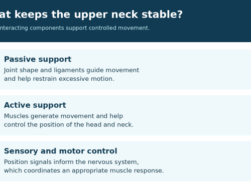
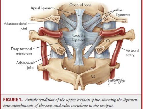
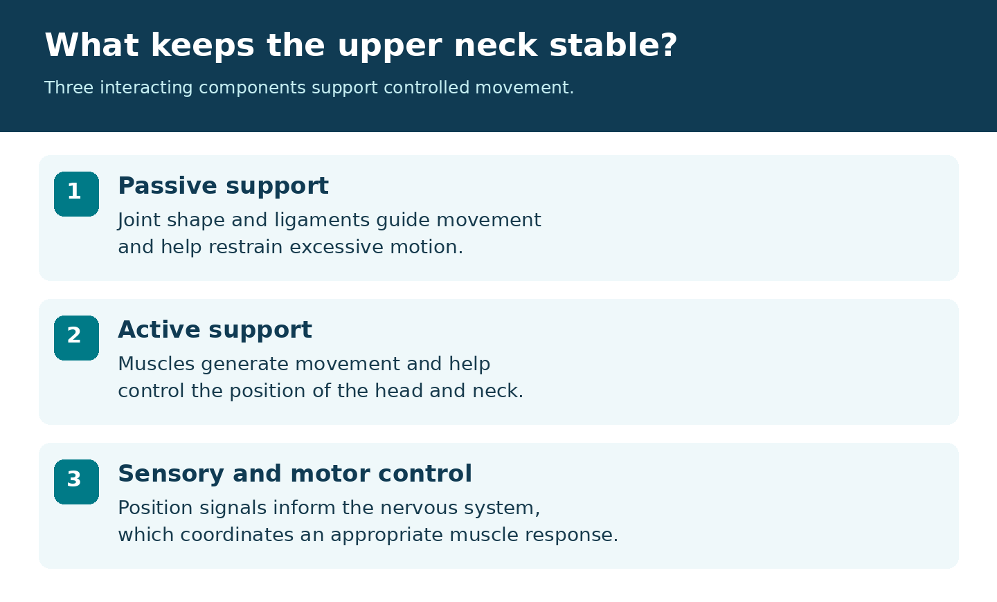
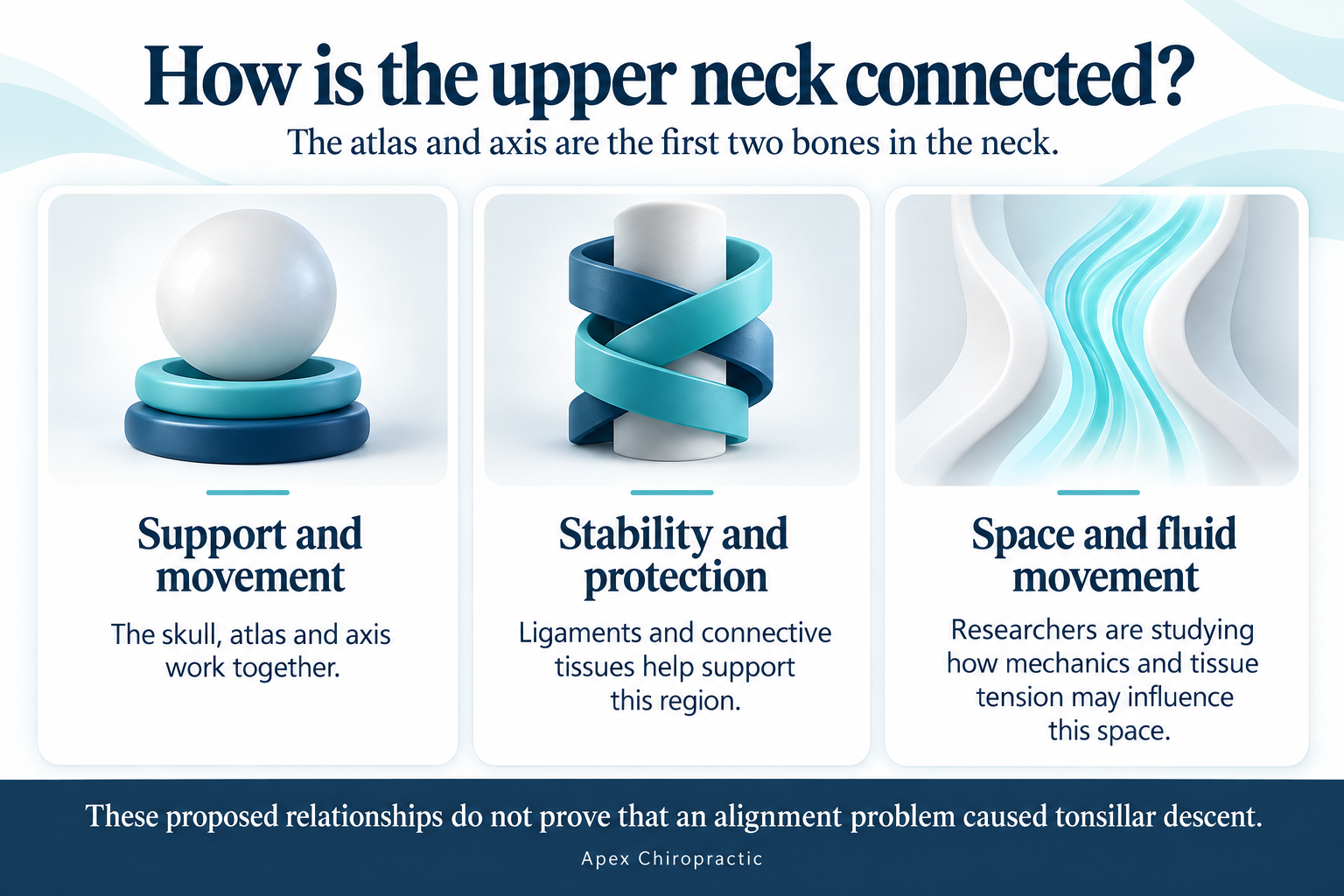
The skull, atlas, and axis work together to support the head and allow movement. The atlas and axis are the first two bones in the neck. Ligaments and other connective tissues help stabilize this region. Nearby tissues also support and surround the brainstem and spinal cord. Researchers have proposed that injury or altered upper cervical mechanics may influence tension on these tissues and affect the space available for fluid movement.[2] These proposed relationships are one reason to consider the upper neck during a thorough evaluation. They do not mean that every person with tonsillar ectopia has an alignment problem or that an alignment problem caused the descent.
Can Forward Head Posture Make Symptoms Worse?
Forward head posture means the head rests farther in front of the shoulders. It often combines bending through the lower neck with extension near the base of the skull to keep the eyes looking ahead. Sustained positioning can add mechanical demands to the neck muscles and supporting tissues.
Posture, Balance, and Autonomic Function
The relationship may involve more than muscle strain. Research discussed in the DCCJP clinical neurology materials has examined forward head posture in relation to sensorimotor control and autonomic nervous system function. Sensorimotor control helps coordinate information about head position, eye movements, and balance. The autonomic nervous system helps regulate automatic functions such as heart rate, blood pressure, and sweating.
A 2020 study compared 80 people with forward head posture with 80 people of similar age, sex, and body mass index who had normal head alignment. The forward head posture group showed differences in eye tracking during neck rotation, balance, and accuracy when repositioning the head. The groups also differed in the strength of the skin sympathetic response, an electrical skin response related to sympathetic activity, although its timing did not differ significantly.[6] This supports an association between posture and the measured functions. It does not establish that posture caused the differences or that every autonomic function was impaired.
A 2021 randomized trial examined 110 athletes with chronic neck pain, reduced cervical lordosis, and forward head translation. Both groups received a rehabilitation program, while one group also received a specific cervical traction intervention. The added intervention produced greater improvements in alignment, pain, disability, sensorimotor control, and skin sympathetic response measures.[7] These findings apply to the population and treatment studied, rather than demonstrating that every cervical adjustment produces the same effects.
Why Might Neck Position Matter?
One proposed explanation is altered sensory input from the neck. Neck muscles and joints send information to the nervous system about head position and movement. Changes in that input may influence the way the brain coordinates balance, eye movements, and autonomic responses. This is a proposed mechanism, rather than proof that forward head posture directly compresses the vagus nerve or causes dysautonomia.
For a person with cerebellar tonsillar ectopia, a coexisting neck problem could add to the overall symptom burden. However, these studies did not establish that forward head posture causes tonsillar descent, obstructs CSF flow, or enlarges a syrinx. Posture and autonomic findings should be considered alongside the neurological examination and imaging.
Comfortable screen positioning, regular changes of position, and individually selected rehabilitation may help address a separate neck concern. Care should account for a syrinx or suspected instability. New weakness, worsening balance, or swallowing changes require medical assessment rather than being attributed to posture.
What Symptoms Can Occur with Cerebellar Tonsillar Ectopia?
Many people with low lying tonsils have no symptoms. When symptoms are present, they may involve several areas.
Headaches and Neck Pain
A characteristic Chiari headache occurs at the back of the head or near the base of the skull. It may become worse with coughing, sneezing, laughing, lifting, or straining. These activities briefly change pressure within the head and spinal canal. Neck pain or tightness near the base of the skull may also occur. However, migraine and other common causes of headache can coexist and need separate consideration.
Dizziness and Balance Difficulties
Some patients experience dizziness, unsteadiness, or difficulty walking. Others describe a rocking sensation or sensitivity to movement. These symptoms can also arise from inner ear problems, migraine, and other conditions. They should not automatically be blamed on the MRI finding.
Visual Changes
Possible complaints include blurred vision, double vision, or the sensation that the surroundings are moving. Abnormal eye movements may also be identified during an examination.
Swallowing, Voice, or Breathing Changes
Difficulty swallowing, choking, hoarseness, or breathing problems during sleep may occur when nearby neurological structures are affected. These symptoms warrant medical assessment.
Changes in Sensation or Strength
Symptoms may include:
- Numbness or tingling
- Burning sensations
- Changes in pain or temperature sensation
- Hand weakness
- Difficulty with buttons or other precise movements
- Dropping objects
- Changes in walking or coordination
Some of these symptoms are particularly important when a syrinx is present.
Symptoms must be interpreted alongside the examination and imaging because many conditions can produce similar complaints.
Clinical Examination for Cerebellar Tonsillar Ectopia
An MRI shows structure. An examination helps determine how the nervous system is functioning.
Depending on your symptoms, an evaluation may assess:
- Muscle strength and hand coordination
- Sensation
- Reflexes
- Eye movements
- Cranial nerve function
- Balance and walking
- Coordination of the arms and legs
The pattern of findings matters. Changes that are progressing over time deserve particular attention.
Evaluating Cerebellar Tonsillar Ectopia on MRI
MRI is the primary imaging examination for evaluating the cerebellar tonsils and surrounding structures. The review includes more than the amount of descent.
Important questions include:
- How much space remains around the tonsils and brainstem?
- Are the fluid spaces open or crowded?
- Is there pressure on the brainstem or spinal cord?
- Is a syrinx present?
- Are there other abnormalities at the base of the skull or upper neck?
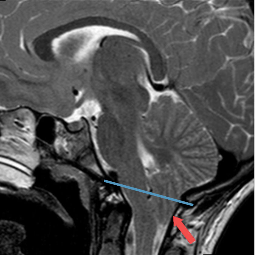
Tonsillar position is measured using McRae’s line, which connects the front and back edges of the foramen magnum. Images from the side, front, and across the region can help provide a more complete picture. The two tonsils may not sit at the same height, making it important to review more than one image.[2]
Upright MRI for Cerebellar Tonsillar Ectopia
Most MRI examinations are performed while lying down. Upright imaging allows clinicians to examine anatomy while the person is seated or standing. In selected situations, this may provide additional information about the effect of posture. Research has reported differences in the frequency of detected tonsillar ectopia between certain upright and lying down patient groups.[3] This does not mean everyone needs upright imaging or that a conventional MRI is inadequate. The appropriate examination depends on the clinical question.
Cine MRI and CSF Flow in Cerebellar Tonsillar Ectopia
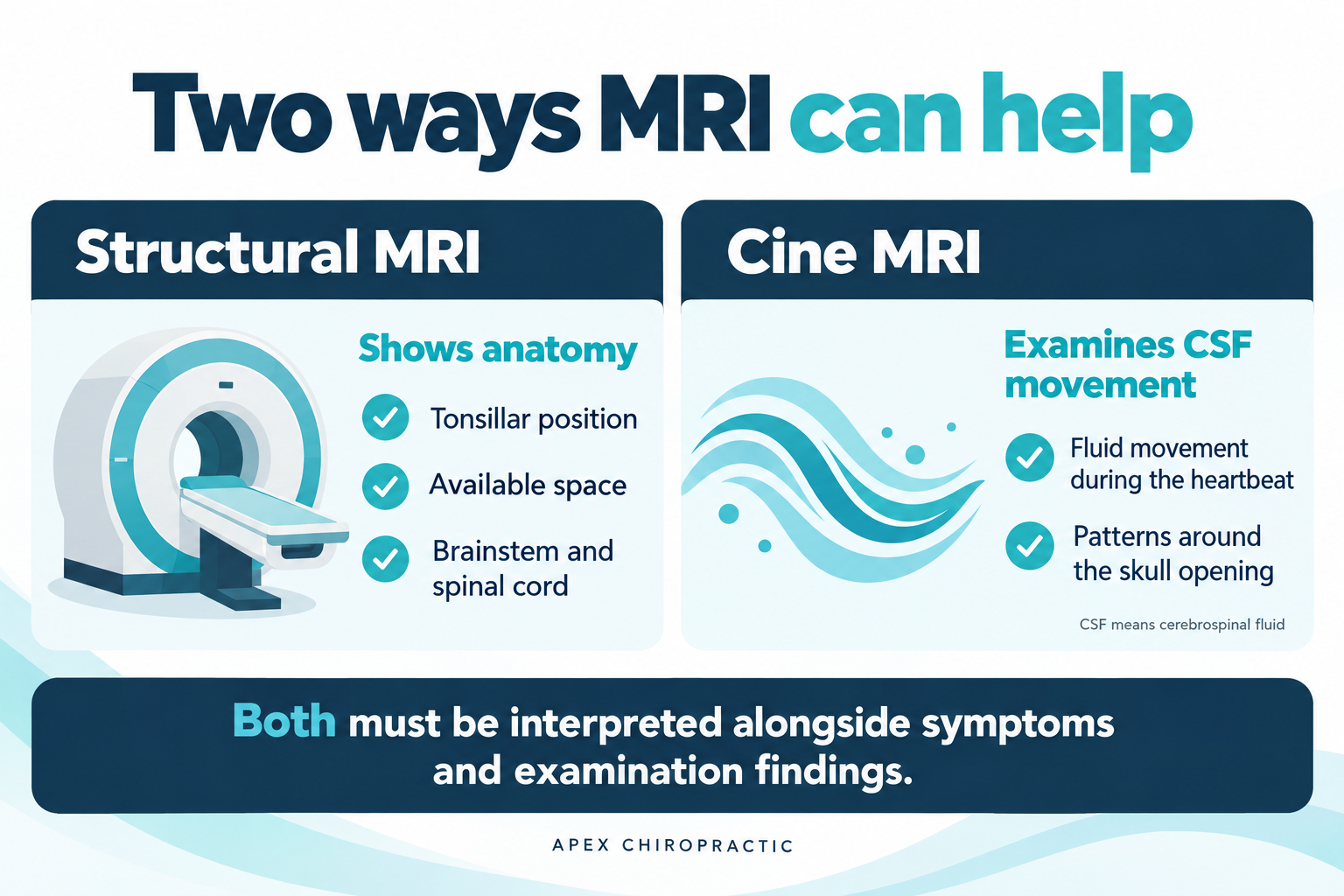
A phase contrast cine MRI evaluates cerebrospinal fluid movement during the heartbeat. A regular MRI helps show where structures are located. Cine MRI adds information about how fluid moves around them. It may help clarify whether crowding is affecting fluid movement. Its results must still be interpreted with the rest of the examination and imaging.
What Are the Treatment Options for Cerebellar Tonsillar Ectopia?
Treatment depends on the complete clinical picture.
Monitoring Cerebellar Tonsillar Ectopia
An incidental finding without concerning symptoms or associated abnormalities may not require treatment. A clinician may recommend observation, follow up examinations, or additional imaging depending on the circumstances.
Managing Symptoms of Cerebellar Tonsillar Ectopia
Some patients benefit from treatment directed at headaches or a separate neck or balance problem. Activity modification may also be appropriate when certain movements or straining consistently provoke symptoms. These approaches should be tailored to the person and should not delay evaluation of neurological changes.
What Is Chiari 0?
Chiari 0, sometimes called Chiari zero, describes a condition in which crowding or restricted cerebrospinal fluid (CSF) movement at the base of the skull may cause problems despite little or no downward displacement of the cerebellar tonsils. Originally, the term described patients who had syringomyelia, a fluid filled cavity within the spinal cord, without tonsillar herniation, and whose syrinx improved after posterior fossa decompression.[4]
Some more recent publications describe Chiari 0 with or without a syrinx. Under this broader definition, the absence of a visible spinal cord cavity does not necessarily exclude the condition. Clinicians consider symptoms, neurological findings, crowding, and evidence of disturbed CSF movement together.[5]
However, the diagnostic criteria are still evolving. Headaches, dizziness, or mildly low lying tonsils alone do not establish Chiari 0. Careful specialist assessment helps determine whether imaging findings explain the symptoms and whether other conditions should be considered. MRI and, in selected cases, cine MRI may help guide that evaluation. Chiari 0 reinforces an important point: the available space and fluid movement can matter as much as the measurement of tonsillar descent.
Where Does Upper Cervical Chiropractic Fit?
An upper cervical evaluation may help identify a separate neck concern and determine whether further medical assessment is appropriate. Published material describes changes in CSF flow imaging following a specific form of image guided atlas adjustment.[2] These observations are an area of investigation. They do not establish that all upper cervical techniques produce the same effects or that chiropractic care reliably reverses tonsillar descent or resolves a syrinx. Any proposed care of the neck must take the neurological examination and imaging findings into account. Suspected instability, pressure on the brainstem or spinal cord, a syrinx, or progressive neurological symptoms should combine specialist assessment along with cervical adjustments.
When Is a Surgical Consultation Appropriate?
A neurological or neurosurgical consultation may be appropriate when there is:
- Progressive weakness or other neurological decline
- A symptomatic or enlarging syrinx
- Pressure on the brainstem or spinal cord
- Persistent, disabling Chiari type headaches
- Significant disruption of CSF movement
A consultation does not automatically mean surgery is needed. When surgery is appropriate, posterior fossa decompression creates more space around the opening at the base of the skull. Some procedures also expand the protective covering around the brain and spinal cord. The approach depends on the individual findings.
When Should You Seek Urgent Care?
New or rapidly worsening weakness, substantial difficulty walking, or worsening swallowing problems requires prompt medical assessment. Sudden severe headache, new symptoms suggesting a stroke, or difficulty breathing requires emergency care.
Understanding Cerebellar Tonsillar Ectopia in Context
The most useful question is not simply, “How many millimeters have my tonsils descended?”
It is: “Does this finding fit my symptoms, and is it affecting nearby structures or fluid movement?”
At Apex Chiropractic in Neptune Beach, an evaluation can include discussion of upper cervical concerns and whether additional neurological or neurosurgical assessment is appropriate. Bring your MRI report and, when available, the actual images. Understanding the complete picture helps guide the next step.
This article provides general education and does not replace an individualized medical evaluation.
References
- Milhorat TH, Chou MW, Trinidad EM, et al. Chiari I malformation redefined: Clinical and radiographic findings for 364 symptomatic patients. Neurosurgery. 1999;44(5):1005 to 1017.
- Rosa S, Baird JW, Harshfield D, Chehrenama M. Craniocervical Junction Syndrome: Anatomy of the Craniocervical and Atlantoaxial Junctions and the Effect of Misalignment on Cerebrospinal Fluid Flow. In: Hydrocephalus: Water on the Brain. 2018. DOI: 10.5772/intechopen.72890.
- Freeman MD, Rosa S, Harshfield D, et al. A case control study of cerebellar tonsillar ectopia (Chiari) and head and neck trauma (whiplash). Brain Injury. 2010;24(7 to 8):988 to 994. DOI: 10.3109/02699052.2010.490512.
- Iskandar BJ, Hedlund GL, Grabb PA, Oakes WJ. The resolution of syringohydromyelia without hindbrain herniation after posterior fossa decompression. Journal of Neurosurgery. 1998;89(2):212–216. DOI: 10.3171/jns.1998.89.2.0212.
- Saadeh A, et al. Chiari zero malformation with syringobulbia. Radiology Case Reports. 2023. Full text.
- Moustafa IM, Youssef A, Ahbouch A, Tamim M, Harrison DE. Is forward head posture relevant to autonomic nervous system function and cervical sensorimotor control? Cross sectional study. Gait & Posture. 2020;77:29–35.
- Moustafa I, Youssef ASA, Ahbouch A, Harrison D. Demonstration of Autonomic Nervous Function and Cervical Sensorimotor Control After Cervical Lordosis Rehabilitation: A Randomized Controlled Trial. Journal of Athletic Training. 2021;56(4):427–436.