By William Soriano, DC | Upper Cervical Chiropractor | Apex Chiropractic
The craniocervical junction of an infant or young child should not be viewed simply as a smaller version of the adult upper cervical spine. During growth and development, the relationship between the skull, atlas (C1), axis (C2), joints, muscles, nervous system, and stabilizing ligaments continues to change. Incomplete ossification, different skeletal proportions, increased mobility, ligamentous laxity, and developing neuromuscular control all contribute to biomechanics that differ significantly from those of an adult and influence the functional environment of the alar ligaments in children.
Among the important stabilizing structures of this region are the alar ligaments in children, paired ligaments located deep within the craniocervical junction. Understanding these ligaments in babies and children requires understanding not only the ligaments themselves, but the developing system in which they function.
What Are the Alar Ligaments?
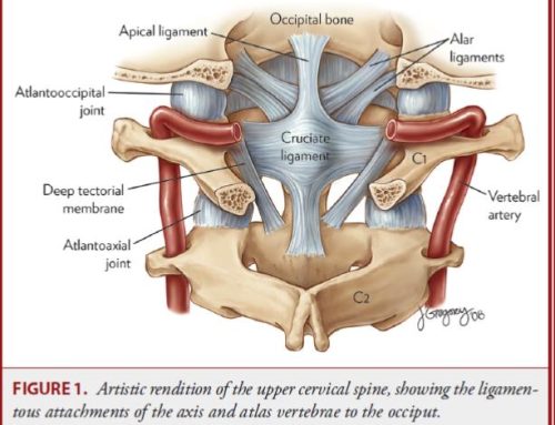
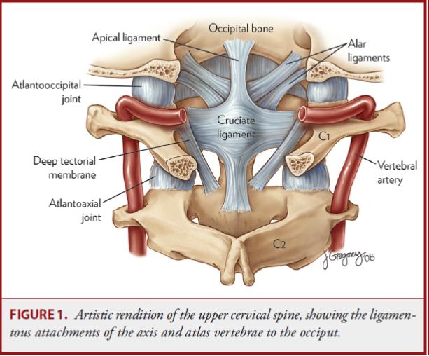
The alar ligaments are strong paired structures located deep within the craniocervical junction. They extend primarily from the upper lateral aspects of the dens, or odontoid process of C2, toward the medial region of the occipital condyles at the base of the skull. Anatomical variation in their orientation and morphology is well documented. Together with structures such as the transverse ligament of the atlas, tectorial membrane, joint capsules, osseous anatomy, and surrounding musculature, the alar ligaments contribute to stabilization of the junction between the head and upper cervical spine. Their biomechanical importance is particularly associated with restraining excessive upper cervical movement, including axial rotation. For a more detailed discussion of adult anatomy, biomechanics, injury mechanisms, MRI findings, and clinical examination, see our comprehensive guide to the alar ligaments.
This distinction is important because the alar ligaments do not stabilize the head and neck by themselves. They function as one component of an integrated craniocervical stabilization system.
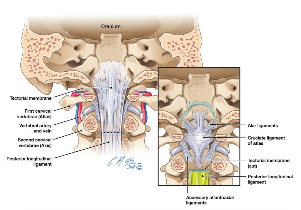
Figure 1. Ligamentous anatomy of the craniocervical junction. Removal of the tectorial membrane reveals the deeper alar and cruciate ligaments connecting and stabilizing the occiput, atlas (C1), and axis (C2).
The Pediatric Craniocervical Junction Is Not a Small Adult Spine
Several anatomical and biomechanical characteristics distinguish the infant and young child’s cervical spine from the adult cervical spine.
These include:
- A proportionally larger and heavier head relative to the body
- Greater ligamentous laxity
- Developing cervical musculature
- Incomplete ossification
- Open synchondroses
- More horizontally oriented facet joints
- Greater overall cervical mobility
- Developing neuromuscular and postural control
These differences have significant biomechanical consequences. Younger children demonstrate different patterns of cervical motion and injury than adults, with pediatric cervical instability and traumatic injuries showing particular importance at the upper cervical spine.
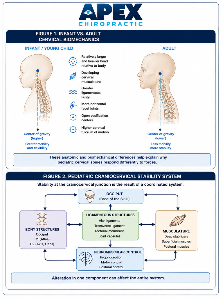
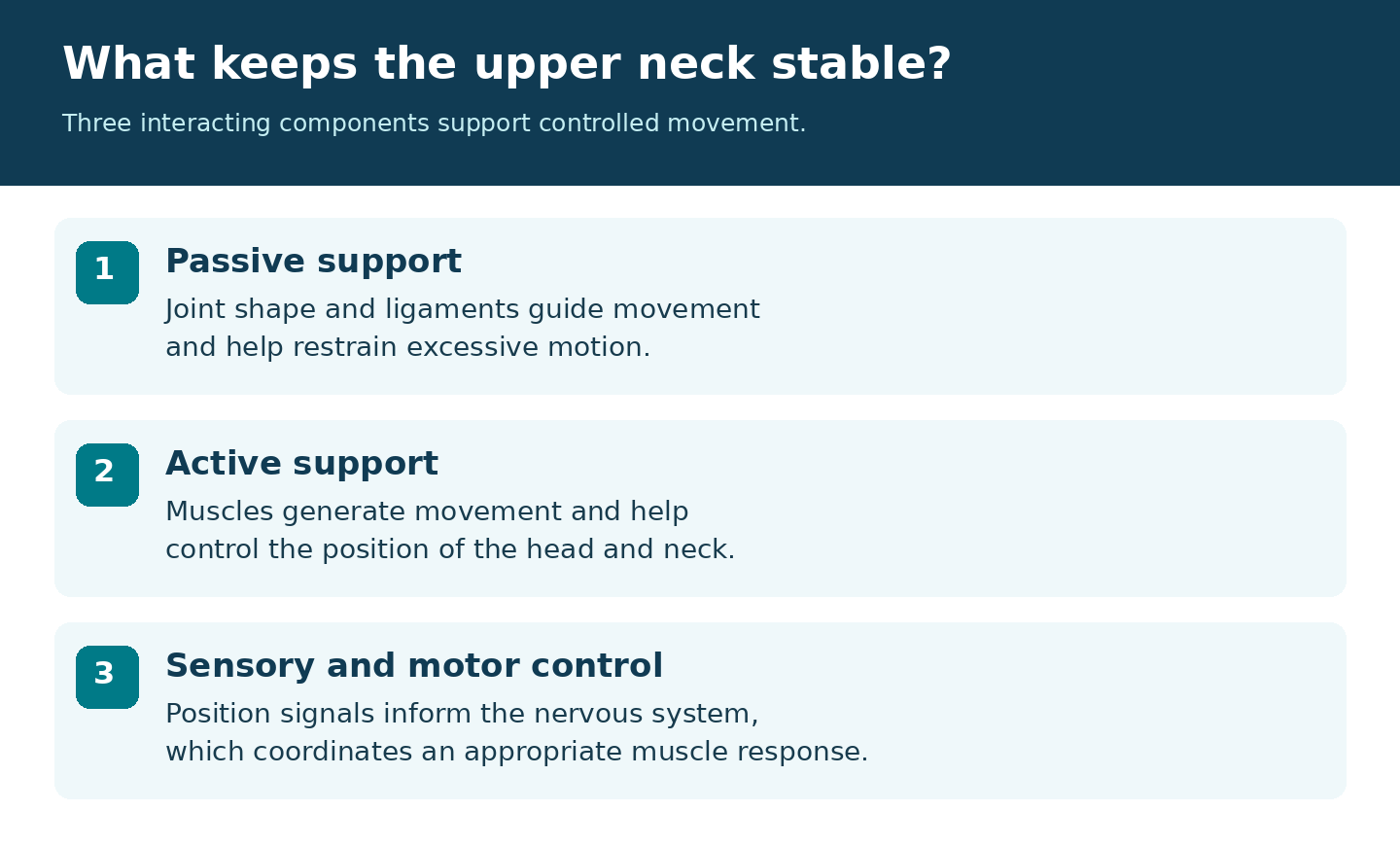
FIGURE 2: The pediatric cervical spine differs substantially from the adult spine in head-to-body proportion, skeletal development, ligamentous laxity, facet orientation, muscular development, and overall mobility. Craniocervical stability depends on the coordinated interaction of bone, ligaments, musculature, and neuromuscular control.
This is one reason adult biomechanical assumptions cannot simply be applied to infants.
Development of C1 and C2
The atlas and axis undergo substantial development throughout childhood. At birth, C1 and C2 have multiple ossification centers separated by cartilaginous synchondroses. These structures progressively ossify and fuse as the child grows. Understanding this normal developmental anatomy is essential because immature anatomy can sometimes resemble fracture, instability, or other pathology when interpreted using adult criteria.
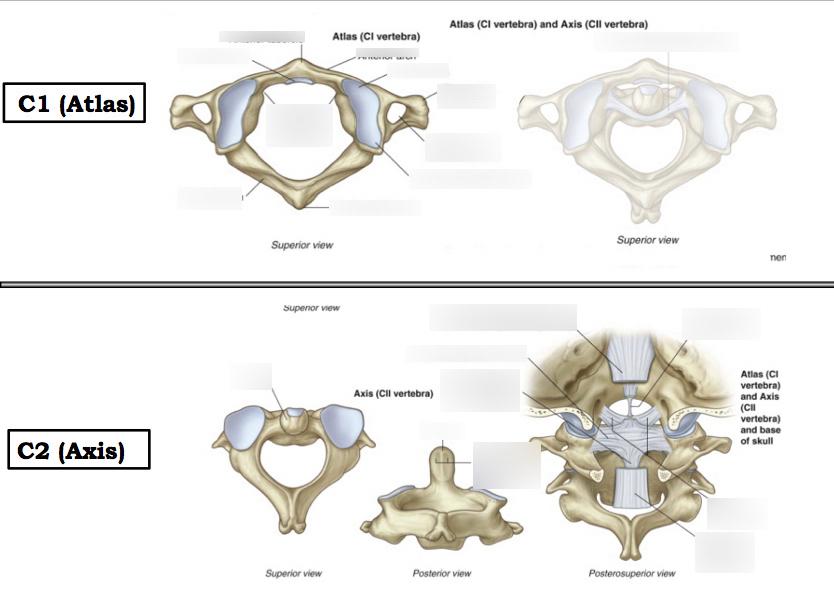
Figure 3. Anatomy of the atlas (C1) and axis (C2), the two uppermost cervical vertebrae. Their unique architecture permits substantial motion at the craniocervical junction and provides attachment sites for its major stabilizing ligaments.
The development of the craniovertebral junction itself is complex. Embryologically, the occiput, atlas, axis, dens, and associated structures arise through segmentation and resegmentation of specialized embryonic tissues. Developmental abnormalities within this process can produce clinically significant congenital anomalies of the craniocervical junction.
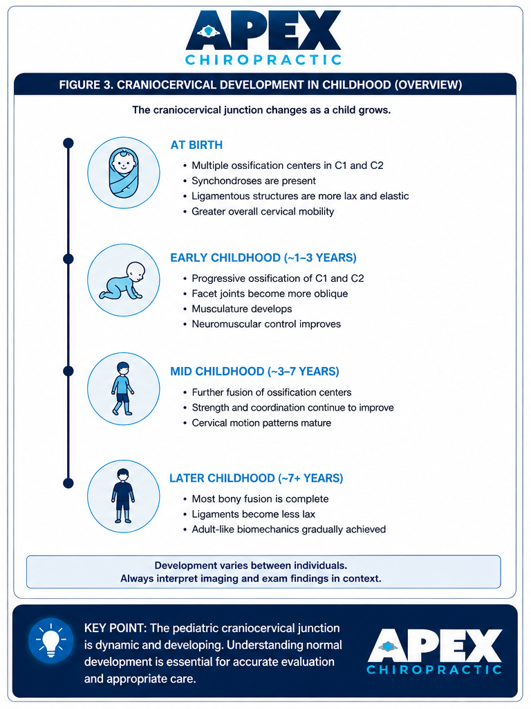
Figure 4. The craniocervical junction continues to mature throughout childhood. Progressive ossification, changing joint anatomy, muscular development, and improving neuromuscular control gradually produce more adult-like cervical biomechanics.
This developmental anatomy is one reason pediatric upper cervical imaging must be interpreted according to the child’s age rather than adult standards.
The Alar Ligaments and Motion Control
The upper cervical spine allows substantial movement between the head, atlas, and axis. The alar ligaments in children participate in controlling that motion. Their orientation allows them to become tensioned during particular combinations of upper cervical movement, contributing to restraint of excessive motion.
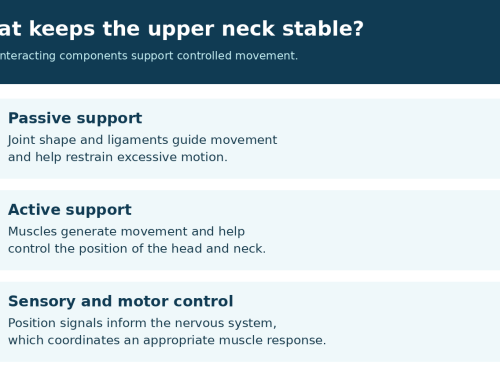
However, normal stability depends upon the coordinated interaction of multiple systems:
Ligamentous structures
- Alar ligaments
- Transverse ligament
- Tectorial membrane
- Joint capsules
Bony structures
- Occiput
- Atlas (C1)
- Axis (C2)
- Dens
Muscular structures
- Deep cervical stabilizers
- Suboccipital musculature
- Superficial cervical muscles
Neuromuscular control
- Proprioception
- Motor control
- Postural control
The pediatric craniocervical junction should therefore be thought of as a dynamic stabilization system, rather than simply a collection of individual ligaments.
What About Birth?
During childbirth, an infant’s head and cervical spine may experience combinations of rotation, flexion, extension, compression, and traction. The specific mechanical environment varies substantially depending upon fetal position, maternal anatomy, duration and progression of labor, method of delivery, and whether assistance or instrumentation is required.
At the same time, the infant cervical spine has anatomical characteristics that differ considerably from those of an adult. That makes the biomechanics of birth an important area of consideration when evaluating an infant.
However, an important scientific distinction must be made. The presence of mechanical forces during delivery does not by itself establish that the alar ligaments were injured. The available literature does not establish that routine birth or even a mechanically difficult delivery commonly causes alar ligament injury.
Likewise, feeding difficulty, head preference, irritability, cranial asymmetry, or torticollis following birth should not automatically be interpreted as evidence of an alar ligament injury. These findings have numerous possible causes and require appropriate clinical evaluation.
Can the Alar Ligaments Be Injured in Children?
Yes. Actual traumatic injuries involving the alar ligaments in children have been documented. Caird and colleagues reported three pediatric/adolescent patients with persistent neck pain and torticollis following trauma in whom isolated alar ligament disruption was identified.
A separate case report documented a child with traumatic atlantoaxial rotatory dislocation in whom rupture of an alar ligament was demonstrated with MRI. More recent literature continues to describe isolated unilateral alar-ligament injuries and emphasizes that these injuries are uncommon and that diagnosis and management remain challenging because of the limited number of reported cases.
These cases establish an important point: Pediatric alar-ligament injury is real, but it should not be presumed from symptoms alone.
Alar Ligament Injury vs. Altered Upper Cervical Function
This distinction is particularly important clinically. A child may have altered cervical movement, muscular asymmetry, head-position preference, torticollis, discomfort, or other findings without having a torn alar ligament. Conversely, significant ligamentous injury may occur in traumatic circumstances and may require advanced imaging and specialist evaluation. Therefore, altered upper cervical biomechanics does not equal alar ligament injury. The two concepts should not be used interchangeably.
How Strong Are a Child’s Alar Ligaments?
At present, there is insufficient data to establish a specific tensile failure threshold for the alar ligaments of a living infant or young child. This is important because biomechanical studies of adult cadaveric alar ligaments are sometimes used to describe ligament strength. Those values should not be directly extrapolated to babies or children. Pediatric tissues are developing, the surrounding anatomy is immature, and the mechanical behavior of the entire craniocervical complex differs from that of an adult.
Imaging the Alar Ligaments in Children
Pediatric upper cervical imaging requires knowledge of normal developmental anatomy. Normal pediatric findings can potentially be mistaken for pathology because of differences involving:
- Ossification centers
- Synchondroses
- Atlantodental relationships
- Vertebral morphology
- Cervical alignment
- Joint relationships
- Prevertebral soft tissues
A detailed understanding of developmental anatomy is therefore essential when evaluating the pediatric craniocervical junction. CT provides excellent visualization of osseous anatomy. MRI provides superior evaluation of soft tissues and can visualize important ligamentous structures of the craniocervical junction.
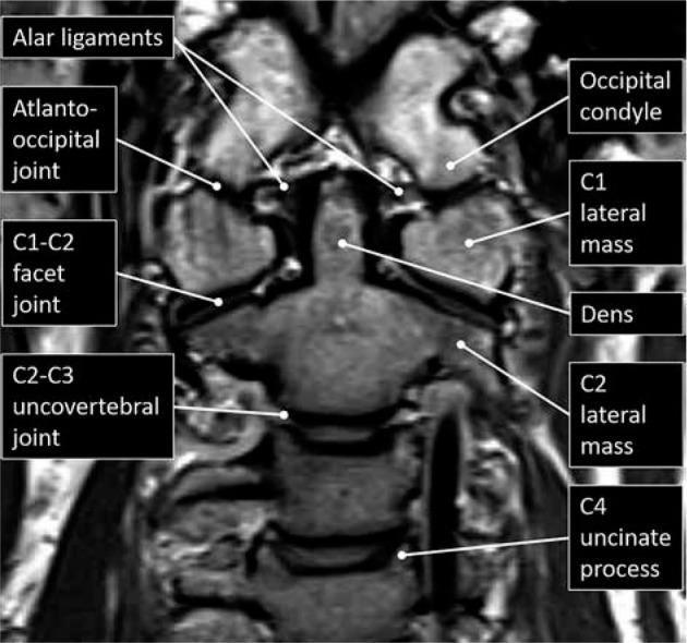
Figure 5. Coronal MRI demonstrating the alar ligaments and their relationship to the dens, occipital condyles, C1 lateral masses, and upper cervical articulations.
When significant ligamentous injury is suspected, imaging findings must be interpreted together with the child’s age, mechanism of injury, symptoms, neurological findings, physical examination, and developmental anatomy.
Babies With Torticollis, Head Preference, or Feeding Difficulties
Infants may present with concerns such as:
- Persistent head rotation or head preference
- Torticollis
- Difficulty comfortably turning the head in one direction
- Cranial asymmetry
- Breastfeeding or bottle-feeding difficulties
- Difficulty maintaining certain feeding positions
- History of a difficult or instrument-assisted delivery
These presentations warrant thoughtful examination of the infant’s neuromusculoskeletal system. The cervical spine and craniocervical junction may appropriately be considered as part of that examination. However, these findings should not automatically be attributed to an injured alar ligament. The documented pediatric cases of alar-ligament disruption involve traumatic injuries and should not be generalized to infants presenting with common developmental, positional, or feeding concerns. That distinction is essential to responsible clinical interpretation.
Why This Matters Clinically
Understanding pediatric craniocervical anatomy serves two equally important purposes. First, it helps clinicians understand normal development, movement, and biomechanics. Second, it helps identify circumstances that may require additional medical evaluation.
Significant trauma, neurological abnormalities, suspected instability, fracture, ligamentous disruption, or other red-flag findings warrant appropriate medical investigation and, when indicated, advanced imaging or specialist referral. For conservative clinicians working with pediatric patients, recognizing when a presentation requires medical investigation or specialist referral is just as important as understanding what may appropriately fall within the scope of conservative care.
The Bigger Picture
The alar ligaments are fascinating structures, but they are only one component of an extraordinarily complex developing system. Throughout childhood, the relationship among the skull, C1, C2, ligaments, joint capsules, muscles, sensory systems, and nervous system continues to mature.
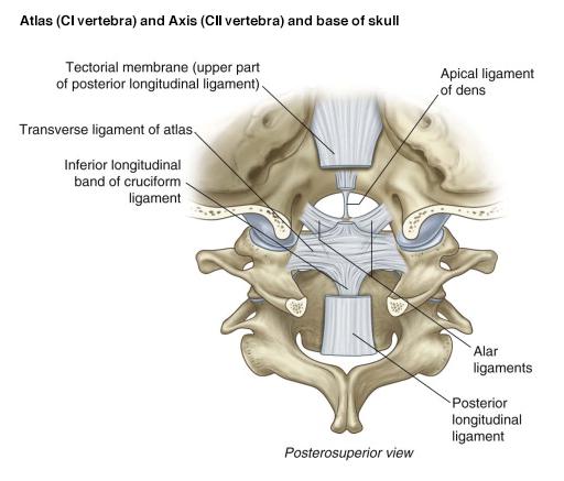
Figure 6. Posteriosuperior view of the craniocervical junction demonstrating the alar ligaments in relation to the occiput, atlas (C1), axis (C2), transverse ligament, cruciform ligament, and tectorial membrane.
That is perhaps the most important concept to take away: the pediatric craniocervical junction is dynamic and developing. Understanding normal development gives clinicians a better framework for evaluating pediatric upper cervical function while helping prevent adult anatomical assumptions from being incorrectly applied to babies and children.
When alar ligament injury is suspected, the diagnosis should be based on the mechanism of injury, clinical presentation, appropriate imaging, and evaluation of the entire craniocervical system and not on a single symptom or finding.
References
- Ghanem I, El Hage S, Rachkidi R, Kharrat K, Dagher F, Kreichati G. Pediatric cervical spine instability. Journal of Children’s Orthopaedics. 2008;2(2):71–84. doi:10.1007/s11832-008-0092-2. PubMed | Free Full Text (PMC)
- Lustrin ES, Karakas SP, Ortiz AO, Cinnamon J, Castillo M, Vaheesan K, et al. Pediatric cervical spine: normal anatomy, variants, and trauma. RadioGraphics. 2003;23(3):539–560. doi:10.1148/rg.233025121. PubMed | Publisher / DOI
- Menezes AH. Craniocervical developmental anatomy and its implications. Child’s Nervous System. 2008;24(10):1109–1122. doi:10.1007/s00381-008-0600-1. PubMed
- Pang D, Thompson DNP. Embryology and bony malformations of the craniovertebral junction. Child’s Nervous System. 2011;27(4):523–564. doi:10.1007/s00381-010-1358-9. PubMed | Free Full Text (PMC)
- Gopinathan NR, Viswanathan VK, Crawford AH. Cervical spine evaluation in pediatric trauma: a review and an update of current concepts. Indian Journal of Orthopaedics. 2018;52(5):489–500. doi:10.4103/ortho.IJOrtho_607_17. PubMed | Free Full Text (PMC)
- Smoker WRK, Khanna G. Imaging the craniocervical junction. Child’s Nervous System. 2008;24(10):1123–1145. doi:10.1007/s00381-008-0601-0. PubMed
- Caird MS, Hensinger RN, Vander Have KL, Gelbke MK, Farley FA. Isolated alar ligament disruption in children and adolescents as a cause of persistent torticollis and neck pain after injury: a report of three cases. Journal of Bone and Joint Surgery American. 2009;91(11):2713–2718. doi:10.2106/JBJS.H.01405. PubMed
- Niibayashi H. Atlantoaxial rotatory dislocation: a case report. Spine. 1998;23(13):1494–1496. doi:10.1097/00007632-199807010-00012. PubMed
- Iwanaga J, Sardi J, Voin V, Chapman JR, Oskouian RJ, Tubbs RS. Anatomy of alar ligament Part I: morphometrics and variants. World Neurosurgery. 2017;107:1001–1006. doi:10.1016/j.wneu.2017.07.187. PubMed
- Reeves BC, Valcarce-Aspegren M, Robert SM, Elsamadicy AA, Tucker A, Storm PB, DiLuna ML, Kundishora AJ. Isolated unilateral alar ligamentous injury: illustrative cases. Journal of Neurosurgery: Case Lessons. 2024;7(14):CASE23664. doi:10.3171/CASE23664. PubMed | Free Full Text (PMC)